Grover PS, Lorton L The incidence of unerupted permanent teeth and related clinical cases. Oral Surg Oral Med Oral Pathol. 1985; 59:420-425
Raghoebar GM, Boering G, Vissink A, Stegenga B Eruption disturbances of permanent molar: a review. J Oral Pathol Med. 1991; 20:159-166
Raghoebar GM, Boering G, Jansen HWB, Vissink A Secondary retention of permanent molar: a histologic study. J Oral Pathol Med. 1989; 18:427-431
Proffit WR, Vig KW Primary failure of eruption: a possible cause of posterior open-bite. Am J Orthod. 1981; 80:173-190
Oliver RG, Richmond S, Hunter B Submerged permanent molars: four case reports. Br Dent J. 1986; 160:128-130
Brearley LJ, McKibben DH Ankylosis of primary teeth: 1. prevalence and characteristics. ASDC J Dent Child. 1973; 40:54-63
Jalevik B, Moller M Evaluation of spontaneous space closure and development of permanent dentition after extraction of hypomineralized permanent first molars. Int J Paediatr Dent. 2007; 17:328-335
Mullally BH, Blakeley D, Burden DJ Ankylosis: an orthodontic problem with a restorative solution. Br Dent J. 1995; 179:1126-1130
This report describes the prosthetic management of a 15-year-old patient with an infra-occluded first permanent molar due to primary failure of eruption (secondary retention). An indirect composite onlay restoration was used to stimulate the periodontal fibres, improve function and restore occlusal stability. This paper describes the clinical technique involved.
CPD/Clinical Relevance: Early detection, diagnosis and management of infra-occluded permanent molar teeth is important to avoid occlusal complications, in addition to improving function and stimulating the periodontal fibres.
Article
Several terms can be used to describe a tooth with arrested eruption and an abnormal position below the occlusal plane, such as infra-occlusion or submergence.
Generalized arrested or delayed dental eruption is usually related to a syndrome or systemic condition (Table 1). On occasion, one permanent molar tooth may have arrested or delayed eruption, which is not associated with a syndrome or systemic condition. There is a prevalence of 0.01% in first permanent molars, and 0.06% in second permanent molars.1 For diagnostic purposes, affected teeth can be classified (Table 2) as:
Impaction;
Primary retention; or
Secondary retention.
Syndromic
Non-Syndromic
Apert syndrome
Cleidocranial dysplasia
Crouzon syndrome
Endocrine disorders
Ectodermal dysplasia
Gardner syndrome
HIV infection
Pfieffer syndrome
Sclerosteosis
Tricho-dento-osseous syndrome
Abnormal position or deformity of tooth germ
Ankylosis
Gingival fibromatosis or hyperplasia
Lack of space
Localize bone deformity, such as cleft palate
Mechanical obstruction
- Supernumerary tooth
- Cyst
- Odontogenic or non-odontogenic tumour
Primary Failure of Eruption (PFE)
- Primary retention
- Secondary retention
Impaction
Cessation of eruption of a tooth caused by a clinically or radiographically detectable physiological barrier in the eruption path, or due to an abnormal position of the tooth.2
Primary retention
Cessation of eruption of a normally placed and normally developed tooth before gingival emergence without a recognizable physical barrier in the eruption path, and when the tooth is delayed more than two years.2
Secondary retention
Cessation of eruption of a tooth after emergence without a physical barrier or ectopic position of the tooth.3
Primary failure of eruption (PFE) is defined as non-syndromic eruption failure of permanent teeth in the absence of mechanical obstructions. This condition demonstrates some or all of the features listed below, as described by Proffit and Vig:4
Posterior teeth are commonly involved;
It can occur in both primary and permanent molar teeth;
Involvement may be unilateral or bilateral;
Involved teeth erupt into initial occlusion and then cease to erupt further (secondary retention), or may fail to erupt entirely (primary retention);
Involved teeth become ankylosed; and
Involved teeth cannot be moved by application of an orthodontic force.
As PFE incorporates features of both primary and secondary retention, it suggests that this condition may have two separate mechanisms or two manifestations of the same mechanism.5
The dental follicle is responsible for bone resorption during intra-osseous dental eruption. Primary retention is most likely caused by a disturbance of the dental follicle that fails to initiate the metabolic events responsible for bone resorption.6
The aetiology of secondary retention is unknown, however, a possible role of ankylosis has been suggested.2 The two reliable signs of ankylosis are infra-occlusion and immobility; only one-third of cases exhibit a metallic sound on percussion or radiographic obliteration of the periodontal ligament space.3 A retrospective diagnosis of ankylosis can be made when a tooth does not respond to orthodontic forces.
Consequences of infra-occlusion of a permanent molar tooth, dependant upon severity (Table 3), can include:
Tilting of adjacent teeth;
Overeruption of opposing tooth;
Occlusal instability and interferences;
Dental midline shift;
Lack of development of the involved alveolar area; and
Increased risk of dental caries and/or localized gingival inflammation, secondary to difficult access for plaque control.
Mild
Occlusal surface of infra-occluded tooth is 1 mm below the occlusal plane.
Moderate
Occlusal surface of infra-occluded tooth is approximately level with the contact point of one or both adjacent tooth surfaces.
Severe
Occlusal surface of infra-occluded tooth is level with or below the interproximal gingival tissue of one or both adjacent tooth surfaces.
Management
While the condition may be clinically apparent, diagnosis can be confirmed by referral to a paediatric dentist or orthodontist. Multidisciplinary management is aimed to stimulate the periodontal fibres, improve function and restore occlusal stability.
The treatment options for infra-occluded first permanent molars are:
No treatment;
Surgical removal; and
Direct or indirect composite build-up.
No treatment
No treatment is most appropriate in cases of mild infra-occlusion, and would require regular monitoring by the general dental practitioner.
Surgical removal
Surgical removal is indicated if infra-occlusion of the first permanent molar is detected early, so that removal at the ideal age range (8–10 years)7 would lead to spontaneous development and space closure. However, this is seldom the case, as the affected tooth may erupt into initial contact before cessation of eruption. Therefore, most infra-occluded permanent molars are detected beyond this age range.
Surgical removal in the teenage patient would result in an edentulous space and bony defect, and precipitate occlusal instability. This would have implications if orthodontic space closure or future osseointegrated implant placement were planned. In addition, it carries the risk of damage to the inferior alveolar nerve and adjacent structures, and fracture of the mandible.
Direct or indirect composite build-up
Prosthetic management ensures occlusal stability, and allows preservation of alveolar bone until after the pubertal growth spurt (9–15 years of age in females and 11–17 years of age in males)8 for osseointegrated implant placement. This avoids disturbance of normal development of the jawbones.
Composite resin is the preferred material as it is adhesive, conservative of tooth tissue, reversible, tooth-coloured and easily adjusted or repaired. It can be placed as a direct or indirect (without tooth preparation) restoration. The indirect approach is able to reproduce ideal contours, morphology and contacts with adjacent and opposing teeth that might be difficult to achieve with large, direct composite build-up restorations.
Prosthetic management is only possible in some cases, dependant upon certain selection criteria (Table 4). It was a viable option for the patient described in this case report.
Affected tooth has a good prognosis and is not: grossly carious, heavily restored, worn down or hypomineralized
Beyond the ideal age range (8-10 years) for extraction of first permanent molar
Case report
A 15-year-old patient was referred to the Paediatric Dental Unit at St George's Hospital (London) by his orthodontist regarding his infra-occluded lower left first permanent molar (LL6). The patient reported some difficulty cleaning the affected tooth. He was accompanied by his father and did not report any family history of similar eruption disturbances. His general dental practitioner provided routine care. He was motivated and had no habits such as thumb-sucking or parafunction. Medical history was clear.
Extra-oral examination
Nothing abnormal detected;
Skeletal Class I;
Average vertical skeletal proportions.
Intra-oral examination
Nothing abnormal detected with soft tissues. Oral hygiene was moderate with BPE score of 1 in all sextants.
All permanent teeth (excluding permanent third molars) had erupted into the mouth, without any signs of dental caries, enamel hypomineralization or tooth surface loss. The upper and lower arches were well aligned, with Class I incisor relationship and Class I molar relationship on the right side. The dental midlines were both central and coincident. Canine guidance was observed on lateral excursion on both left and right sides, and no occlusal interferences were detected.
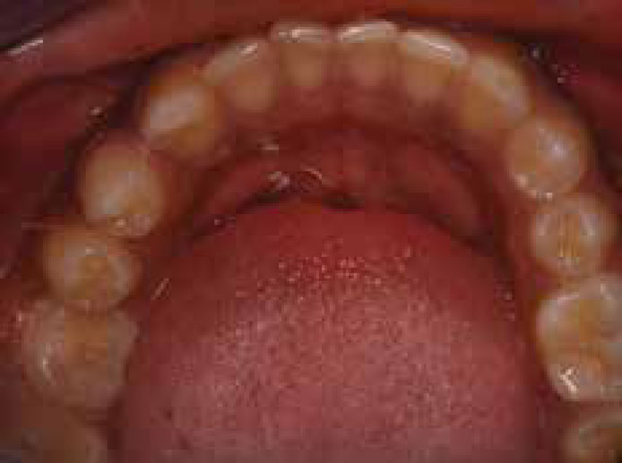
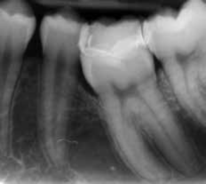
Infra-occlusion of LL6 was evident on examination (Figure 1); vertical distance of 4 mm from occlusal surface to occlusal plane. In addition to infra-occlusion, the following features suggested ankylosis of LL6:
It was immobile;
It exhibited a high-pitched metallic sound on percussion; and
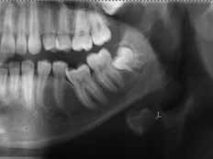
Radiographically, partial obliteration of the periodontal ligament space was evident (Figure 2).
There was slight tilting of adjacent teeth, but no overeruption of the opposing teeth. Radiographic examination (Figure 2) revealed completed root development, good bone levels, no caries, no periapical pathology and the presence of unerupted permanent third molar teeth. LL6 responded positively to ethyl chloride and electric pulp vitality tests.
Diagnosis
Infra-occlusion of the lower left first permanent molar (LL6) due to primary failure of eruption (secondary retention).
Treatment planning
Treatment planning was carried out in conjunction with an orthodontic consultant. The treatment options were discussed with the patient and his father, along with associated risks and benefits. A decision was made for an indirect composite build-up. This would prevent any further tilting of adjacent teeth. The patient was informed that oral hygiene would need improvement before any treatment is provided.
They were aware that, in the future, the tooth may require surgical removal and prosthetic replacement.
Treatment
On the first treatment visit, an impression of the lower arch was recorded with Extrude (Kerr UK Ltd, Peterborough) and Extrude Extra (Kerr), heavy (Type 1) and light (Type 3) bodied polyvinylsiloxane impression materials, respectively, in a stock tray using a one-stage technique. An alginate impression of the upper arch, to obtain an opposing cast for articulation, followed. Both impressions were sent to the laboratory along with the recorded shade and instructions to fabricate an indirect Gradia (GC Europe NV, Leuven, Belgium), a light-cured micro-ceramic-composite onlay, to bring LL6 into occlusion.
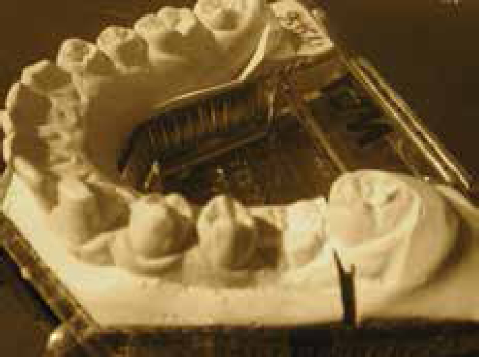
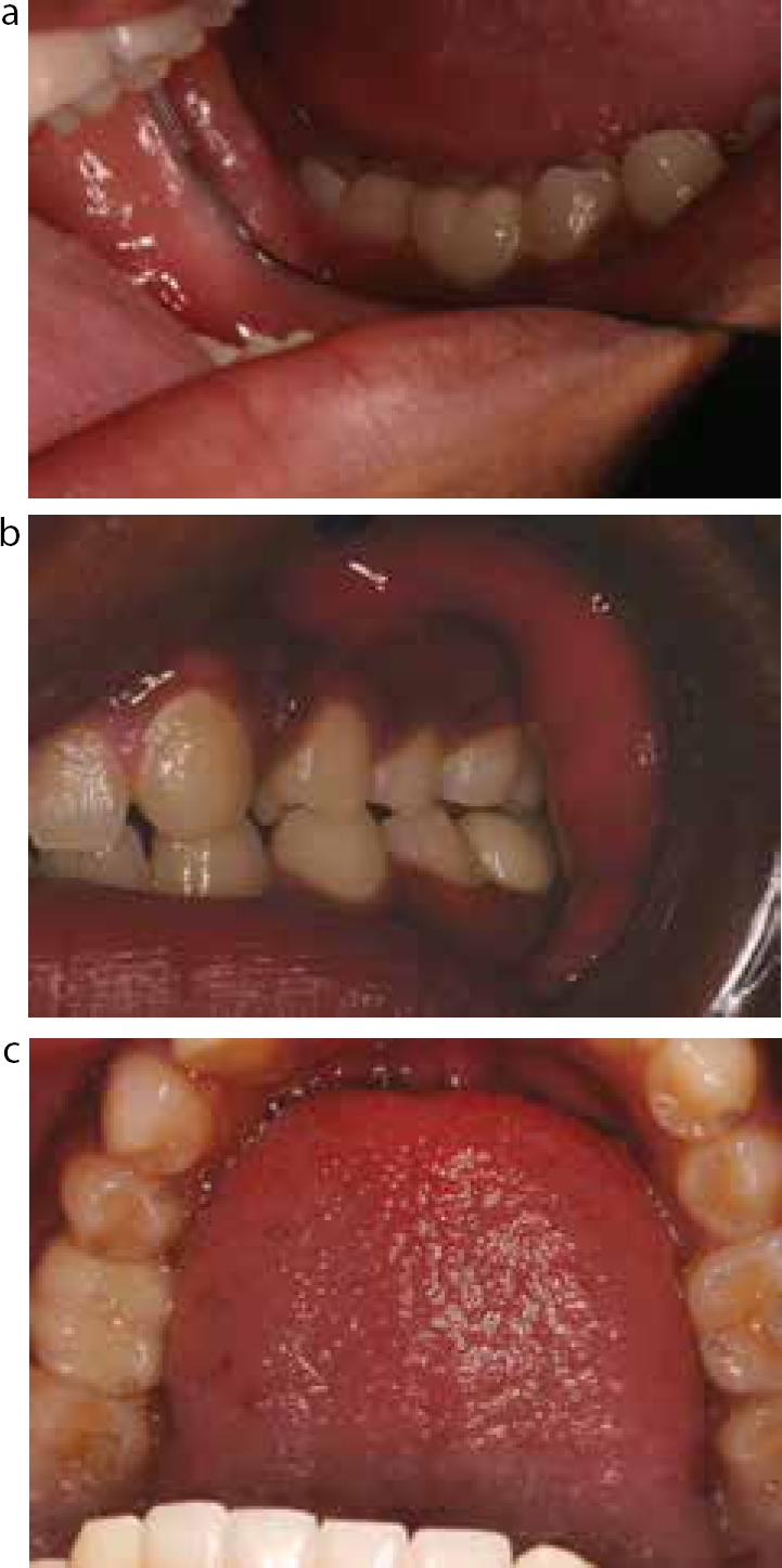
The laboratory provided a dental cast (Figure 3) and composite onlay restoration, which was inspected for fit surface discrepancies, before try-in and cementation. Nexus 3 (Kerr), a resin composite luting cement, was selected. Due to its hydrophobic nature, excellent moisture control is necessary to achieve optimal bonding. For this purpose, rubber dam was used to isolate LL5, LL6 and LL7 (clamp placed on LL7) in a ‘split-dam’ arrangement. Try-in gel was applied to the occlusal surface of LL6, before seating the onlay. The fit, margins, contact points, occlusion and shade match were checked to ensure that no adjustments were necessary. Prior to application of silane primer, the occlusal surface of LL6 was roughened with air abrasion, to enhance micro-mechanical bonding; 37% phosphoric acid gel was used to etch the enamel and dentine. Optibond Solo Plus (Kerr), a total-etch bonding agent, was used according to manufacturer's instructions. A dual-cure NX3 cement was applied onto the onlay and seated firmly; excess cement and contact points were cleared before all surfaces were light-cured. The occlusion of the finished restoration (Figure 4) was checked with articulating paper.
Figure 3. Mandibular dental cast showing infra-occluded LL6, spacing between premolars and mild distal tilting of LL5.Figure 4. Post-cementation (a) left buccal and (b) left oblique and (c) mandibular arch views showing composite onlay on LL6.
The need to maintain good oral hygiene was reinforced.
Review
The patient was reviewed at 4 weeks, 6 months and 1 year (and ongoing) following cementation. The patient did not report any problems. Clinical and radiographic examination (Figure 5), at 1 year post-operatively, showed satisfactory marginal adaptation and no changes in the periodontal tissues. Overall, the patient was satisfied with the aesthetic and functional result.
Figure 5. One year post-cementation radiograph (LCPA) showing satisfactory marginal adaptation and restored interproximal and occlusal contacts of LL6 with adjacent and opposing teeth.
Conclusion
This case highlights the principles of prosthetic management of an infra-occluded permanent molar tooth. Early detection by the general dental practitioner, and diagnosis and treatment planning by a multidisciplinary team, is important to avoid dento-alveolar complications and for a successful outcome. It has been shown that infra-occluded teeth can be brought into functional occlusion, using a non-invasive prosthetic approach. However, this treatment option is not always most appropriate and, therefore, cases must be selected carefully. It is important that oral hygiene is maintained to a high level and periodic monitoring is carried out, to ensure longevity of the restoration.