Fortier LA. Stem cells, classifications, controversies and clinical applications. Vet Surg. 2005; 34:415-423
Chandra Mouli PE, Manoj Kumar S, Senthil B Stem cells in dentistry – a review. J Pharm Sci Res. 2012; 4:(7)1872-1876
Kawaguchi H, Hirachi A, Hasegawa N Enhancement of periodontal tissue regeneration by transplantation of bone marrow mesenchymal stem cells. J Periodontol. 2004; 75:(9)1281-1287
Gronthos S, Brahim J, Li W Stem cell properties of human dental pulp stem cells. J Dent Res. 2002; 81:531-535
Huang A, Chen Y, Chan A. Isolation and characterization of human dental pulp stem/stromal cells from nonextracted crown fractured teeth requiring root canal therapy. J Endod. 2009; 35:673-816
Miura M, Gronthos S, Zhao M, Lu B, Fisher LW, Robey PG SHED: Stem cells from human exfoliated deciduous teeth. Proc Natl Acad Sci. 2003; 100:5807-5812
Sonoyama W, Liu Y, Yamaza T, Tuan RS, Wang S, Shi S Characterization of the apical papilla and its residing stem cells from human immature permanent teeth: a pilot study. J Endod. 2008; 34:166-171
Seo BM, Miura M, Gronthos S, Bartold PM, Batouli S, Brahim J Investigation of multipotent postnatal stem cells from human periodontal ligament. Lancet. 2004; 364:149-155
Morsczeck C, Götz W, Schierholz J, Zeilhofer F, Kühn U, Möhl C Isolation of precursor cells (PCs) from human dental follicle of wisdom teeth. Matrix Biol. 2005; 24:155-165
Nakashima M, Iohara K, Ishikawa M Stimulation of reparative dentin formation by ex vivo gene therapy using dental pulp stem cells electrotransfected with growth/differentiation factor11 (Gdf11). Hum Gene Ther. 2004; 15:1045-1053
Iohara K, Zheng L, Ito M, Tomokiyo A, Matsushita K, Nakashima M. Side population cells isolated from porcine dental pulp tissue with self-renewal and multipotency for dentinogenesis, chondrogenesis, adipogenesis, and neurogenesis. Stem Cells. 2006; 24:2493-2503
Andreasen JO, Borum MK, Jacobsen HL, Andreasen FM. Replantation of 400 avulsed permanent incisors. 1. Diagnosis of healing complications. Endod Dent Traumatol. 1995; 11:51-58
Andreasen JO, Borum MK, Jacobsen HL, Andreasen FM. Replantation of 400 avulsed permanent incisors. 2. Factors related to pulpal healing. Endod Dent Traumatol. 1995; 11:59-68
Kling M, Cvek M, Mejare I. Rate and predictability of pulp revascularization in therapeutically reimplanted permanent incisors. Endod Dent Traumatol. 1986; 2:83-89
Huang GT-J, Sonoyama W, Liu Y, Liu H, Wang S, Shi S. The hidden treasure in apical papilla: the potential role in pulp/dentin regeneration and bioroot engineering. J Endod. 2008; 34:(6)645-651
Hargreaves K, Law A. Regenerative endodontics, 10th edn. In: Hargreaves K, Cohen S (eds). St Louis: Mosby Elsevier; 2011
Iwaya SI, Ikawa M, Kubota M. Revascularization of an immature permanent tooth with apical periodontitis and sinus tract. Dental Traumatol. 2001; 17:185-187
Trope M. Treatment of the immature tooth with a non–vital pulp and apical periodontitis. Dent Clin N Am. 2010; 54:313-324
Thibodeau B, Teixeira F, Yamauchi M, Caplan DJ, Trope M. Pulp revascularization of immature dog teeth with apical periodontitis. J Endod. 2007; 33:680-689
Banchs F, Trope M. Revascularization of immature permanent teeth with apical periodontitis: new treatment protocol?. J Endod. 2004; 30:196-200
Hoshino E, Kurihara-Ando N, Sato I In-vitro antibacterial susceptibility of bacteria taken from infected root dentine to a mixture of ciprofloxacin, metronidazole and minocycline. Int Endod J. 1996; 29:125-130
Hargreaves K, Law A. Regenerative endodontics, 10th edn. In: Hargreaves K, Cohen S (eds). St Louis: Mosby Elsevier; 2011
Petrino J, Boda K, Shambarger S, Bowles W, McClanahan S. Challenges in regenerative endodontics: a case series. J Endod. 2010; 36:536-541
Torabinejad M, Turman M. Revitalization of tooth with necrotic pulp and open apex by using platelet rich plasma: a case report. J Endod. 2011; 37:265-268
Dannan A. Dental-derived stem cells and whole tooth regeneration: an overview. J Clin Med Res. 2009; 1:(2)63-71
Hay MF. The development in vivo and in vitro of the lower incisor and molars of the mouse. Arch Oral Biol. 1961; 3:86-109
Morio I. Recombinant study of the mouse molar cervical loop and dental papilla by renal transplantation. Arch Oral Biol. 1985; 30:557-561
Slavkin HC, Bavetta LA. Odontogenesis in vivo and in xenografts on chick chorio-allantois. I Collagen and hexosamine biosynthesis. Arch Oral Biol. 1968; 13:145-154
Yamada M, Bringas P, Grodin M, MacDougall M, Cummings E, Grimmett J, Weliky B Chemically defined organ culture of embryonic mouse tooth organs: morphogenesis, dentinogenesis and amelogenesis. J Biol Buccale. 1980; 8:127-139
Stem cells are defined as clonogenic, unspecialized cells capable of both self-renewal and multi-lineage differentiation, contributing to regenerating specific tissues. For years, restorative treatments have exploited the lifelong regenerative potential of dental pulp stem cells to give rise to tertiary dentine, which is therapeutically employed for direct and indirect pulp capping. Current applications of stem cells in endodontic research have revealed their potential to continue root development in necrotic immature teeth and transplanted/replanted teeth. Successful application of pulp revascularization is highlighted here with support of a clinical case report. This article also discusses the role of dental stem cells as a promising tool for regeneration of individual tissue types like dentine, pulp and even an entire functional tooth.
CPD/Clinical Relevance: This article will help practising dental surgeons understand the significance of stem cells in dentistry. Clinicians can harness the potential of stem cells using procedures like pulp regeneration/revascularization in endodontics and improve their knowledge on the recent advances in tissue engineering and future applications of dental-derived stem cells.
Article
Stem cells are a unique type of cell that have a specialized capacity for self-renewal and potency, which can give rise to one and sometimes many different cell types. They aid in the replacement of cells that are lost through normal wear, injury or disease.1 Stem cells can be broadly divided into:
Embryonic stem cells; and
Post-natal stem cells.
Post-natal stem cells are multipotent stem cells and have been harvested from different kinds of tissues like bone marrow, umbilical cord, amniotic fluid, brain tissue, liver, pancreas, cornea, dental pulp, and adipose tissue. These stem cells are comparatively easier to isolate, do not have any ethical issues and are commonly used in current day practice.2
Source of stem cells
The oral and maxillofacial region can be treated with stem cells from the following sources:
Bone marrow stem cells (BMSCs);
Stem cells from the oral and maxillofacial region.
Bone marrow stem cells (BMSCs)
Bone marrow stem cells (BMSCs) can be harvested from sternum or iliac crest and are composed of both hematopoietic stem cells and mesenchymal stem cells (MSCs). The majority of oro-maxillofacial oral structures are formed from mesenchymal cells. BMSCs exhibit the ability to generate osteoid and odontoid structures and have demonstrated good ability to form tooth-supporting periodontal structures like cementum, periodontal ligament (PDL) and alveolar bone, suggesting their potential use for treating periodontal diseases.3
Stem cells from oral and maxillofacial region
In the oral and maxillofacial area, different types of dental stem cells have been isolated and characterized and they include:
Dental pulp stem cells (DPSCs);
Stem cells from exfoliated deciduous teeth (SHEDs);
Stem cells from apical papilla (SCAPs);
Periodontal ligament stem cells (PDLSCs);
Dental follicle progenitor cells (DFPCs).
Dental pulp stem cells (DPSCs)
Gronthos first identified post-natal dental pulp stem cells and found that these cells could regenerate a pulp-dentine like complex. These stem cells are found to reside in the central cell rich zone of the pulp.4 An in vitro study by Huang et al demonstrated the ability of DPSCs to form odontoblast-like cells. DPSCs are more easily sourced as they can be obtained from sound teeth that are deemed for extraction for orthodontic or periodontal reasons.5
Stem cells from exfoliated deciduous teeth (SHEDs)
Exfoliating deciduous teeth contain living pulp remnants and are highly proliferative, clonogenic and have multi-differentiation potential. These cells were isolated and characterized by Miura et al.6 SHEDs have advantages over other post-natal stem cells as they are derived from a source which is non-invasive, being naturally disposed and with limited ethical concerns. They have obvious advantages of higher proliferation rate compared with stem cells of permanent teeth, high plasticity and can differentiate into osteoblasts and odontoblasts. Miura et al demonstrated that SHEDs could not differentiate directly into osteoblasts but did induce new bone formation.6
Stem cells from apical papilla (SCAPs)
Stem cells from apical papilla (SCAPs) are a newly discovered population of stem cells by Sonoyama et al.7 In immature teeth, when the roots are still developing, the dental papilla assumes a position apical to the pulp tissue and epithelial diaphragm. This apical papilla is loosely attached to the apex of the root from where it can be easily detached. SCAPs have a higher proliferation rate compared to DPSCs and the capacity to differentiate into functional dentinogenic cells in vivo.7
Periodontal ligament stem cells (PDLSCs)
Periodontal ligament stem cells (PDLSCs) are mesenchymal stem cells obtained from periodontal ligament (PDLSCs) as investigated by Seo et al.8 They are multi-potent cells with similar features to bone marrow stem cells (BMSCs) and DPSCs. These cells are capable of developing different types of tissues such as bone and tooth associated tissues.8
Dental follicle progenitor cells (DFPCs)
Dental follicle progenitor cells (DFPCs) were isolated by Morsczeck et al from follicles of human third molars and, when transplanted into immunodeficient mice, they were able to recreate a new periodontal ligament-like tissue after 4 weeks.9 This has important applications in periodontal engineering.
Understanding the nature of these progenitor/stem cell populations in the oral cavity is important in determining their potentialities for use in regeneration and tissue engineering. Characterization of stem/progenitor cells, and determination of their potentialities in terms of specificity of regenerative response, may help direct new clinical treatment modalities. Such approaches may provide an innovative and novel biologically-based new generation of clinical materials and/or treatments for dental disease.
Stem cells in dentine tissue engineering
The potential of pulp tissue to regenerate lost dentine is well known. Severe injury to dental pulp from either infection or trauma leads to death of odontoblasts, with a limited ability for regeneration. Healing depends on the intensity and duration of the injury, presence of bacteria, and host immunity. Coronally in the tooth, a new generation of odontoblast-like cells develops from recruitment of a progenitor cell population, leading to reparative dentinogenesis, which is a natural reparative response of the pulp-dentine complex. For years, restorative treatments have exploited this lifelong regenerative potential of dental pulp stem cells, to give rise to tertiary dentine, which is therapeutically employed for direct and indirect pulp capping.
The utilization of post-natal stem cells in clinical applications may be best served by developing materials that stimulate migration of stem/progenitor cells to the site of injury. It is tempting to consider the exciting possibilities of clinically directing this aspect of natural regeneration, both by maximizing recruitment of progenitor cells to areas of injury and disease and also through influencing the nature of the cell populations recruited. Although recruitment of progenitor cells to sites of injury occurs naturally, it may be considered random and uncontrolled. Directed recruitment of progenitor cells might be achieved through local application of enriched populations of cells, either by harvesting cells from non-autologous teeth or autologous exfoliated primary teeth.6 Bone morphogenetic proteins (BMPs) play a role in the differentiation of dentine and Growth/differentiation factor 11(Gdf 11) is a novel member of the BMP family. Gdf 11 was expressed in terminally differentiating odontoblasts, implying a role in the differentiation of dental pulp stem cells into odontoblast-like cells.10 BMP-2-treated cultured pulp cells11 and Gdf 11–electrotransfected pulp cells10 (non-viral method of gene transfer to living animals) have been successfully transplanted to surgically amputated pulps, suggesting a possible therapeutic approach to dental regeneration. Interestingly, the initial regenerated tissue exhibits an osteo-dentine-like appearance, similar to that of reparative dentine (atubular) rather than tubular dentine. It is worth considering that this may be of benefit in dentine bridge formation, where an atubular dentine would provide a more effective barrier to bacterial progression during any further carious challenge.12 However, this may not be entirely feasible owing to the difficulties of obtaining adequate sources and volume of autologous cells to reduce any immune response to the cell transplant.12
Stem cells in replantation of teeth
Andreasen et al13,14 and Kling et al15 showed excellent radiographic images of the growth of bone and periodontal ligament into the canal space with arrested root formation following replantation of avulsed maxillary incisors. After re-implantation of an avulsed immature tooth, the open apex allows new tissue to grow into the pulp space quickly. The apical part of a pulp may remain vital and may proliferate coronally, replacing the necrotized portion of the pulp. In most cases, the crown of the tooth is intact and ensures no bacterial penetration into the pulp and hence favours new tissue formation. SCAPs and HERS (Hertwigs epithelial root sheath) appear to be important for continued root development as they stimulate the undifferentiated mesenchymal cells in periradicular tissues to differentiate into odontoblasts that contribute to the formation of new dentine and root maturation.16
Stem cells in pulp revascularization
Irreversible injury to dental pulp of an immature permanent tooth from infection or dental trauma before complete root development poses a clinical challenge. Dentine deposition and root maturation ceases, leaving an open root apex and thin dentinal walls that are prone to fracture. Endodontic treatment may be complicated as the root canal system is difficult to debride fully and the open apex provides no barrier stop for the root-filling material. An alternative approach is to provide treatment under conditions where continued dentine formation is promoted. Thus, there is a continued need to develop biologically-based treatment regimens that offer the potential for continued hard tissue formation of the young permanent tooth with a necrotic root canal system and an incompletely developed root. Revascularization is a regenerative treatment and a biologically-based alternative approach to treat necrotic immature teeth. Unlike apexification and artificial apical barrier techniques, it allows for continuation of root development.17 The use of the term ‘revascularization’ was proposed by Iwaya et al18 to describe the clinical healing of periapical abscesses and continued root formation in immature teeth with non-vital pulps. Revascularization of the pulp space in a necrotic infected tooth with apical periodontitis was considered to be impossible. However, if the canal is effectively disinfected and a scaffold into which new tissue can grow is provided, revascularization should occur as in an avulsed immature tooth.19 Regeneration of tissue into the apex of an immature permanent tooth may come from stem cells already residing in the apical papilla, PDL or alveolar bone. Hence components needed for successful revascularization include absence of intracanal infection, a good coronal seal to prevent reinfection, a physical scaffold to promote cell growth and differentiation, as well as signalling molecules for the growth of stem cells.17 Induction of bleeding and the subsequent formation of a blood clot might serve as a scaffold for the periapical cells, including mesenchymal stem cells to migrate into the root canal, and eventually induce new tissue formation within the space.20 The case below describes successful revascularization in a necrotic immature central incisor using mineral trioxide aggregate (MTA) with a follow-up period of 18 months.
Case report
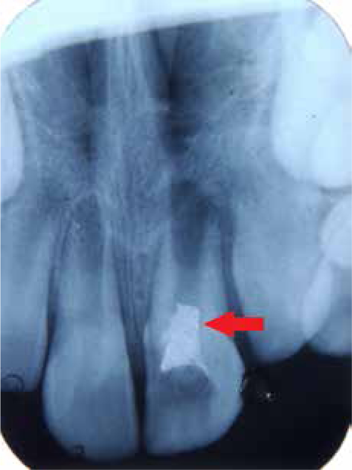
An 8-year-old girl was referred by a general practitioner with a non-vital maxillary left central incisor (UL1) with apical periodontitis, following fracture of a major talon cusp. An MTA pulpotomy was performed to treat pulp exposure of the fractured talon cusp, which had failed. Clinical examination revealed pain on percussion in relation to UL1 and a delayed response to electric pulp testing with no response elicited on using a thermal testing method (cold test). Radiographic examination revealed that the tooth had an incompletely developed apex and periradicular radiolucency (Figure 1). The mother was informed about the limitations and advantages of revascularization as a treatment modality and an informed consent was obtained. The working protocol followed was according to Banchs and Trope,21 to revascularize the pulp in infected necrotic immature roots. The first step was disinfection of the canal. This was achieved by passively irrigating with 20 mL of 5.25% NaOCl for 20 min and then drying with absorbent points. A freshly prepared antibiotic paste consisting of ciprofloxacin, metronidazole and minocycline (100 mg of each drug in a 0.5 mL total volume, glycerine or saline used as vehicle) was placed into the canal using a lentulo spiral to 3 mm short of working length. Most of the revitalization/regeneration procedures use this triple antibiotic paste, also called a Hoshino's paste.22 The access cavity was sealed with Cavitä (3M ESPE, Seefeld, Germany). Instrumentation is contra-indicated in revascularization treatments as root dentinal walls are so thin that any instrumentation makes them weaker and more susceptible to future fractures. At the 3-week follow-up appointment, the patient was asymptomatic, and the tooth showed no tenderness to percussion and palpation. The triple antibiotic paste was removed and the canal was dried with absorbent points. The second step was to create a scaffold for the stem cells to repopulate and continue root formation. For this, the apical tissue in the tooth was nudged using an ISO 70 K-file to induce fresh bleeding, and a blood clot was formed 4 mm apical to the cemento-enamel junction (CEJ). After 15 min, MTA was placed over the blood clot. It is a biocompatible sealing material, which allows the regeneration of new tissue adjacent to it. A wet cotton pellet was placed against the MTA, and the tooth was restored temporarily with Cavitä. On the next day, the temporary restoration was removed, and the MTA set was verified. An approximately 2 mm thick layer of glass ionomer cement was placed over the set MTA cement, and the tooth was restored with composite resin (Figure 2). The fractured talon cusp was reduced and the tooth was restored to normal contour.
Figure 1. Preoperative radiograph showing periapical radiolucency in relation to UL1.Figure 2. Post operative radiograph showing MTA placement 2 mm below the CEJ.
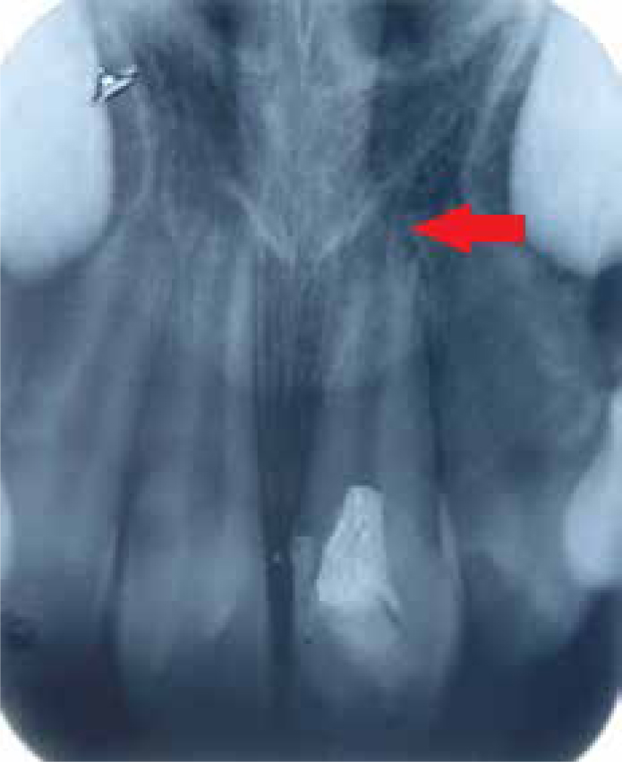
At the 3-month follow-up, the tooth was functional, without sensitivity to percussion and palpation, with normal periodontal findings. At the follow-up examination, the patient continued to be asymptomatic, with a radiographic indication of continued development of the apex of the tooth with complete resolution of the periapical radiolucency. This case had been followed up for 18 months and can be considered successful as root dentinal walls have thickened and the apex has formed normally (Figure 3). Pulp testing (cold, thermal test) did not elicit any response and the tooth remained asymptomatic in all the recall visits.
Figure 3. 18-month follow-up radiograph showing almost complete root development (Nolla stage 9 of root development).
The importance of a bacteria-tight coronal seal for successful revascularization is well documented.23 A few cases have reported positive responses to cold and EPT after a regenerative endodontic procedure.24 The presence or absence of responses in these teeth depends on the coronal level of tissue grown in the root canal and the thickness of filling materials over this tissue. As in the present case, if the filling materials are placed beyond the cemento-enamel level, the tooth is more likely to elicit a negative response to cold or EPT. The presence of a thick layer of restorative material(s) can also prevent stimulation of vital tissues within the root canal.25 Discoloration of the clinical crown is mentioned as a disadvantage in some of the revascularization reports possibly due to the presence of minocycline in the triple antibiotic paste and/or use of grey MTA.24 In this particular case, however, no clinically relevant discoloration was seen, probably due to the use of white MTA and a direct composite restoration. Thibodeau et al reported successful use of cefaclor instead of minocycline in triple antibiotic paste, which might be an effective approach to prevent the discoloration caused by minocycline.20
This method of achieving continued root formation is an example of pulp regeneration and the beginning of stem cell technology in endodontics. It is important to distinguish between the terms ‘revascularization’ and ‘pulp regeneration’. Presently we can only state with certainty that the pulp space has returned to a vital state but, based on research in avulsed teeth and a recent study on infected teeth, it is more likely that the tissue in the pulp space is more similar to periodontal ligament than to pulp tissue. It appears that there is about a 30% chance of pulp tissue re-entering the pulp space.19,20 The predictability of this procedure and the type of tissue that develops in these cases are still to be studied.
Stem cells for bioroot engineering
Dental implants have recently gained momentum as a preferred option for replacing missing teeth instead of bridges or removable dentures. However, the fundamental pitfall is the lack of a natural structural relationship with the alveolar bone (ie the absence of PDL). The alternative approach is to use SCAPs and PDLSCs to form a bioroot. Using a minipig model,26 autologous SCAPs and PDLSCs were loaded onto hydroxyapatite tricalcium phosphate (HA/TCP) and gelfoam scaffolds, respectively, and implanted into sockets of the lower jaw. Three months later, the bioroot was exposed, and a porcelain crown was inserted. This approach is a relatively quick way of creating a root onto which an artificial crown can be installed. The bioroot is different from a natural root in that the root structure is developed by SCAPs in a random manner. Nevertheless, the bioroot is encircled with periodontal ligament tissue and appears to have a natural relationship with the surrounding bone. What remains to be improved is the mechanical strength of the bioroot, which is approximately two thirds of a natural tooth.16 This hybrid strategy of autologous dental stem cell engineering may be applicable to human tooth regeneration.
Regeneration of pulp and dentine
Dental pulp tissue engineering was first tested by Mooney et al.27 Since the isolation and characterization of DPSCs and SHEDs, using these stem cells for dentine/pulp tissue regeneration has drawn great interest. Reparative dentine-like structure is deposited on the dentine surface if DPSCs are seeded onto a human dentine surface and implanted into immuno-compromised mice, suggesting the possibility of forming additional new dentine on existing dentine. Nör has shown that, by seeding SHEDs onto synthetic scaffolds seated in a pulp chamber of a thin tooth slice, odontoblast-like cells can rise from the stem cells and localize against an existing dentine surface after implanting the tooth slice construct into immunocompromised mice.28 These findings shed new light on the possibility of generating pulp and dentine in pulpless canals. However, when implanting cells/scaffolds into root canals that have blood supply only from the apical end, enhanced vascularization is needed in order to support the vitality of the implanted cells in the scaffolds. Recent efforts in developing scaffold systems for tissue engineering have been focusing on creating a system that promotes angiogenesis for the formation of a vascular network. These scaffolds are impregnated with growth factors such as vascular endothelial growth factor (VEGF) and/or platelet-derived growth factor or, further, with the addition of endothelial cells. These approaches are particularly important for pulp tissue engineering.16
If the proposed clinical approach is successful, it implies a fundamental breakthrough in clinical endodontic treatments using cell and tissue engineering therapy.
Stem cells in whole tooth regeneration
As bioengineered tooth crown formation requires the interactions of both dental epithelial cell progenitors and mesenchymal cell progenitors (as in natural tooth formation), the ability to bioengineer a tooth of specified size and shape will depend on the ability first to identify, and then guide, the interactions of both types of cells. Methods to guide the interactions of epithelial and mesenchymal post-natal dental stem cells to form dentine and enamel layers characteristic of natural teeth requires modified scaffold materials and designs. The importance of scaffold materials and design for tissue engineering has long been recognized. Scaffold porosity, biocompatibility and biodegradability, the ability to support cell growth, and use as a controlled gene- and protein-delivery vehicle are all highly significant properties. A variety of hydrophilic polymers have been synthesized that provide cell support and guidance. Importantly, scaffold materials provide a three-dimensional macromolecular structure to guide the final shape of bioengineered tissues. Poly-L-lactic acid and poly lactic co-glycolic acid co-polymers have been used to generate composite scaffolds that degrade within a period of a few weeks up to one year.29 From earlier studies, we know it is possible to regenerate tooth crowns if suitable environments are provided.30,31,32,33,34 These culture sites provide nutrients and oxygen to nurture tooth germs. Thus, several choices exist for cultivating small-sized primordia, such as those of teeth, before they can be implanted into their anatomical sites.
Yen and Sharpe suggested two techniques for tooth regeneration; the first of which involved growing dissociated tooth germs on a tooth-shaped scaffold to result in small, complex, tooth-like structures. The second method included growing epithelial and mesenchymal (stem) cells, either from tooth germs or other sources, in organ culture and, through epithelial-mesenchymal interactions, forming organized teeth.35 Although many challenges remain, stem cell-based tissue engineering of teeth could be a choice for the replacement of missing teeth in the future. A therapeutic option that was unthinkable a few years ago seems an achievable goal today.
Conclusion
We have moved on from the surgical model to the medical model and are likely to move on to the biological model of care with stem cells. Regeneration of the dental tissues provides an attractive alternative to more traditional restorative approaches because the diseased tissue is replaced by natural tissue, which forms an integral part of the tooth. We clearly have an opportunity to move restorative dentistry to a new era, harnessing the biological activity of stem cells and tissue regeneration, but there is still a need to translate research into clinical realities.