The immune system: basis of so much health and disease: 7. antibodies Crispian Scully Eleni A Georgakopoulou Yazan Hassona Dental Update 2024 44:9, 707-709.
The immune system is the body's primary defence mechanism against infections, and disturbances in the system can cause disease if the system fails in defence functions (in immunocompromised people), or if the activity is detrimental to the host (as in auto-immune and auto-inflammatory states). A healthy immune system is also essential to normal health of dental and oral tissues. This series presents the basics for the understanding of the immune system; this article covers antibodies.
Clinical Relevance: Modern dental clinicians need a basic understanding of the immune system as it underlies health and disease.
Article
Crispian Scully Eleni A Georgakopoulou Yazan Hassona
Antibodies (immunoglobulins) are a heterogeneous group of proteins employed by the immune system specifically to identify, neutralize and destroy pathogens.
General properties of antibodies
Antibodies are produced by activated B-lymphocytes (plasma cells) and react specifically with the antigen that stimulated their production; they make up approximately 20% of the plasma proteins. Antibodies confer humoral immunity – protection mainly against pyogenic (pus-producing) bacteria. There are five different classes of antibodies (IgG, IgM, IgD, IgE and IgA); each has a unique function, described below, but the primary function of all antibodies is to bind antigens, and enhance their inactivation and destruction.
Antibody production is usually dependent upon, and modulated by, T-cells. These either assist (T-helper [Th] cells), or moderate (T-suppressor [Ts] cells) antibody production via several cytokines, including interleukins (IL) 1–7, and B-cell growth factor (BCGF).
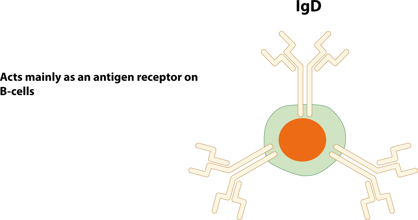
Antibodies can be present both as soluble proteins in the circulation or on the surface of B-cells as membrane-bound proteins known as B-cell receptors (BCR).
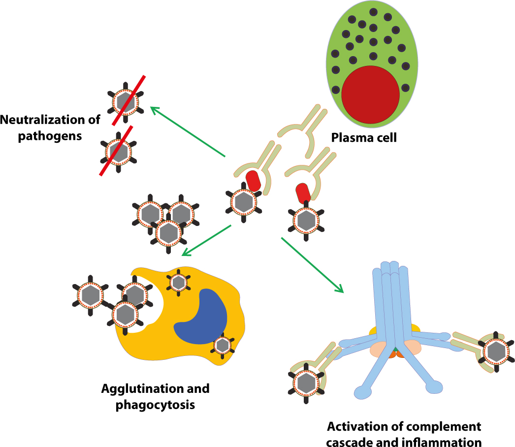
Binding of antibodies to their antigens can result in:
Neutralization: where antibodies block part of the pathogen's surface to render its attack less effective;
Agglutination/opsonization: where antibodies clump pathogens together making them an attractive target for their ingestion by phagocytic leukocytes (macrophages and polymorphonuclear neutrophilic leukocytes [PMNLs]);
Complement activation: where antigen-antibody complexes (immune complexes) activate the complement cascade and result in lysis of the pathogen and activation of inflammation (Figure 1).
Figure 1. Binding of antibodies to their antigens can cause neutralization, agglutination and complement activation.
Antibody structure
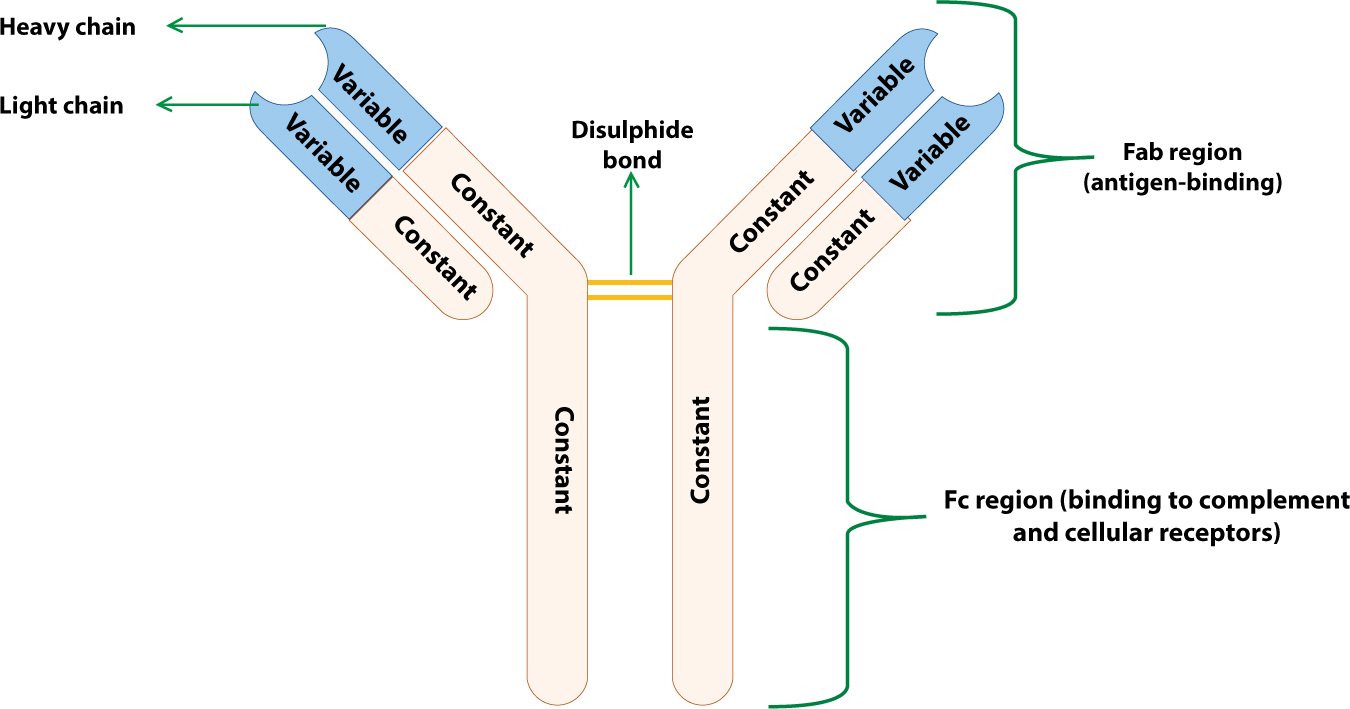
All antibodies have the same basic structure of two identical light (L) chains and two identical heavy (H) chains linked together by disulphide bonds and organized into a Y-shaped structure (Figure 2). The two short arms of the Y-shaped structure consist of light and heavy chains and are able to bind antigens, and thus are termed Fab (fragment antigen-binding). The stem of the Y-shaped structure consists of heavy chains, does not bind antigens but can activate the complement cascade; as it can be crystallized, it is termed Fc (fragment crystallizable) (Figure 2).
Figure 2. Basic antibody structure.
The light and heavy chains are encoded by multi-gene families, and contain regions that are highly variable in their amino acid sequence (termed VL and VH), and regions that have essentially constant amino acid sequences (termed CL and CH) (Figure 2).
The variable regions (V regions) provide antibodies with the diversity required to respond to a vast number of different antibody structures, while the constant regions (C regions) are required for the effector functions of antibodies, such as binding to complement proteins and to receptors on macrophages and other cells.
Antibody classes
Immunoglobulins can be divided into five different classes or isotypes, depending on the structure of their heavy chain. Rearrangement of heavy chain constant (C) region genes generates changes in the immunoglobulin class expressed. There are five different classes of heavy chains with slightly different structures; μ chains for IgM, δ for IgD, ε for IgE, α for IgA and γ for IgG. There are, however, only two forms of the light chains, κ and λ: all immunoglobulin classes can carry either κ or λ, but no single immunoglobulin molecule can have both light chains.
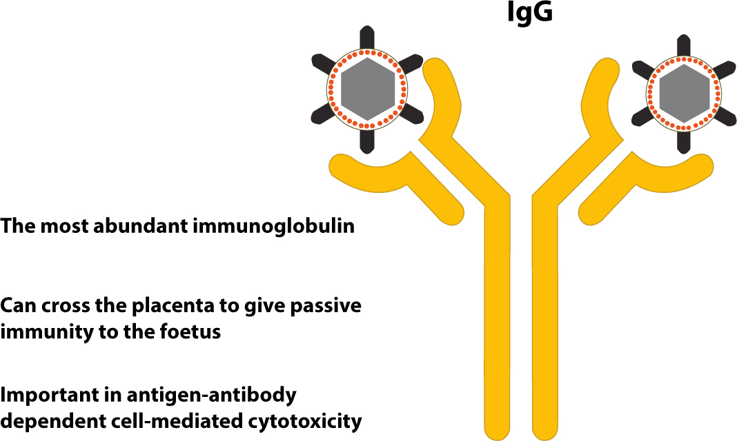
It is the most abundant immunoglobulin class, representing about 80% of serum immunoglobulins;
It is produced predominantly in the secondary immune response, and acts as an opsonin by binding to Fc receptors on phagocytes;
It has four subclasses (IgG1–IgG4). Subclasses IgG1 and IgG3 readily activate complement, whereas IgG2 and IgG4 bind with high affinity to Fc receptors on phagocytes. IgG1, IgG3 and IgG4 cross the placenta, and play a role in protecting the foetus but, since the IgG half life is only about 21 days, the transplacental passage of IgG provides only a temporary immunity.
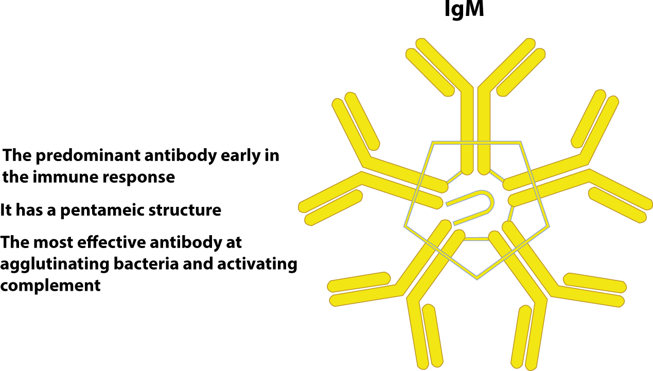
It makes up about 5–10% of total serum immunoglobulins;
It is the major immunoglobulin class produced in the primary immune response;
It is the first to be produced by the neonate, and can readily bind and agglutinate antigens such as bacteria or viruses because it has a pentameric structure;
It activates complement more efficiently than IgG;
It has an important function as a secretory immunoglobulin, but it is too large to cross the placenta or diffuse well into tissue fluids.
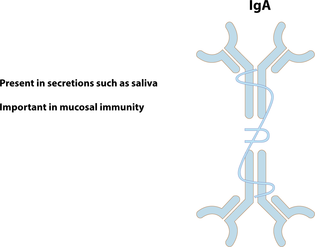
It is the major secretory immunoglobulin, and is present in most external secretions, such as saliva, tears, breast milk, and in the bronchial, digestive and urogenital secretions and mucosae.
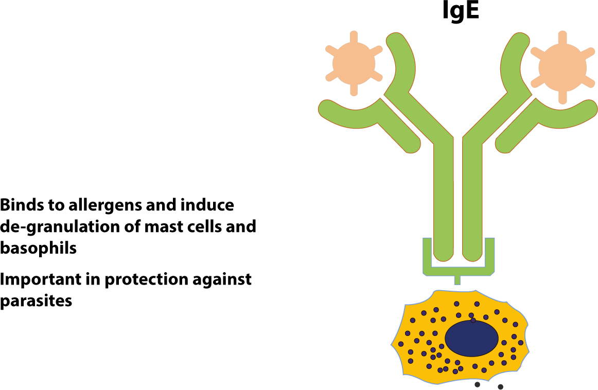
It displays potent biological activity, IgE antibodies binding to Fc receptors on blood basophils and tissue mast cells, causing their de-granulation and release of mediators such as histamine, producing the immediate hypersensitivity reactions responsible for hayfever, asthma and anaphylactic shock.
Figure 6. IgE antibody.
It facilitates the accumulation of cells necessary for local defence against parasites.