Rosetti EP, Marcantonio RAC, Rossa C, Chaves ES, Goissis G, Marcanonio E Treatment of gingival recession: comparative study between subepithelial connective tissue graft and guided tissue regeneration. J Periodontol. 2000; 71:1441-1447
Greene JC, Vermillion JR. The simplified oral hygiene index. J Am Dent Assoc. 1964; 68:7-13
Wang HL, Boyapati L. “PASS” principles for predictable bone regeneration. Implant Dent. 2006; 15:8-17
Tal H. Soft tissue grafts for the treatment of mucogingival deformities and conditions on edentulous ridges. Refu'at Ha-peh Veha-shinayim. 2011; 28:6-16
Zuhr O, Bäumer D, Hürzeler M. The addition of soft tissue replacement grafts in plastic periodontal and implant surgery: critical elements in design and execution. J Clin Periodontol. 2014; 41:S123-S142
Langer L, Langer B. The subepithelial connective tissue graft for treatment of gingival recession. Dent Clin North Am. 1993; 37:243-264
Sedon CL, Breault LG, Covington LL, Bishop BG. The subepithelial connective tissue graft: part II. Histologic healing and clinical root coverage. J Contemp Dent Pract. 2005; 6:139-150
Dual role of subepithelial connective tissue grafting in regeneration of periodontal attachment apparatus Nettemu Sunil Kumar Nettem Sowmya Vijendra Pal Singh Madhu B Verma Dental Update 2024 44:5, 707-709.
Periodontal plastic and aesthetic surgery are gaining significant momentum owing to the increasing aesthetic demands by patients. Along with the fulfilment of aesthetic needs, it is imperative that clinicians also enhance function. From these two important viewpoints, subepithelial connective tissue grafting remains an optimum treatment choice for periodontists when treating gingival recession defects accompanied by periodontal attachment apparatus breakdown.
CPD/Clinical Relevance: Subepithelial connective tissue grafting is a successful procedure in its dual role of gingival recession coverage and predictable periodontal regeneration.
Article
Gingival recession or marginal soft tissue recession is the displacement of the gingival margin apical to the cemento-enamel junction.1 When gingival recession is combined with the presence of pathologically deep periodontal pocket depths and other periodontal parameters, the presentation becomes more complex. This can affect the aesthetic and functional aspects of the periodontal attachment apparatus. When such a combined defect is present, a thorough clinical and radiographic examination, precise diagnosis and meticulous treatment planning is of prime importance. Not all patients are suitable candidates for grafting and regenerative procedures and the clinical decision should be made entirely based on the particular patient diagnosis. Subepithelial connective tissue grafting has been used widely by periodontists in the treatment of gingival recession and as a barrier membrane in guided tissue regeneration therapy owing to its distinct advantages. The subepithelial connective tissue grafts have been highly predictable in gingival recession therapy with respect to a high percentage of root coverage, better healing and less post-operative discomfort at the donor site, when compared to free gingival grafts.2 Also, colour-matching, in relation to the adjacent tissues, offers a clear advantage in comparison to the remaining methods.
This case report highlights the use of subepithelial connective tissue grafting in a dual role whereby root coverage as well as guided tissue regeneration has been achieved.
Case report
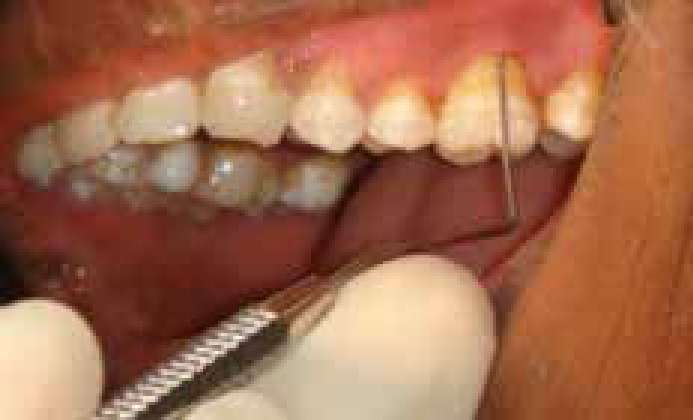
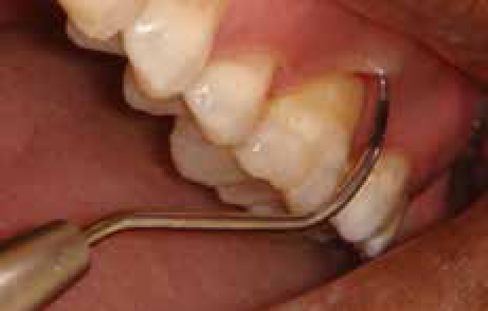
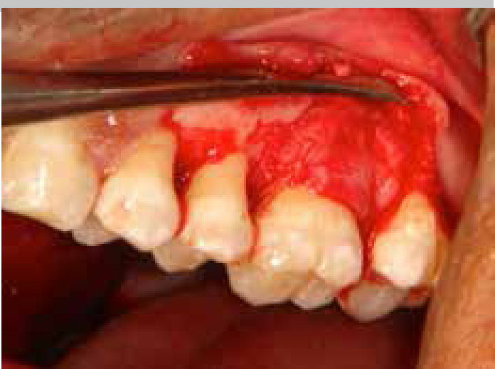
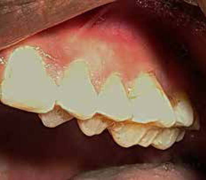
A 28-year-old male patient reported to the Department of Periodontics for a dental check-up. This was his first visit to a dental setting. All histories were non-contributory and the patient was reportedly a teetotaller. Clinical examination showed good oral hygiene according to the Oral Hygiene Simplified Index.3 Examination of UL6 revealed the presence of a Miller‧s Class II gingival recession defect. The recession was around 4 mm between the gingival margin and the cemento-enamel junction as measured using a William‧s graduated periodontal probe (Hu-Friedy Mfg Co, LLC) (Figure 1). There was also Glickman‧s Grade II furcation involvement as evident by the penetration of Naber‧s probe (Hu-Friedy Mfg Co, LLC) into the buccal furcation for a distance of approximately 7 mm (Figure 2). There was no mobility associated with UL6. The remaining dentition was apparently healthy and showed no signs of periodontal involvement. The diagnosis was established as localized aggressive periodontitis in relation to UL6. Interestingly, at this stage, this patient did not show involvement of incisors or other molar teeth and we considered this as an early stage of involvement. The treatment plan for this patient had to take into consideration two important aspects, namely root coverage and periodontal tissue regeneration. Our aim was to minimize patient discomfort and the number of surgical sites, as well as to maximize good results and wound healing. For these reasons, the treatment plan was finalized as subepithelial connective tissue grafting in combination with the use of a xenograft in relation to UL6. Even though an autogenous bone graft is considered as the gold standard in periodontal tissue regeneration, the patient discomfort and healing following the utilization of a third surgical site was taken into consideration. Hence, the use of an osteoconductive xenograft was decided upon. The patient was given a detailed explanation of his periodontal status and the importance of periodontal therapy, combined with a strict maintenance regimen to monitor his periodontal status over a period of time. Written informed consent was obtained from the patient. Phase I therapy included thorough scaling and root planing and a strict maintenance regimen at home.
Figure 1. Pre-operative intra-oral view of a gingival recession defect in UL6.Figure 2. Pre-operative intra-oral view of Glickman‧s Grade II furcation involvement in UL6.
Surgical technique
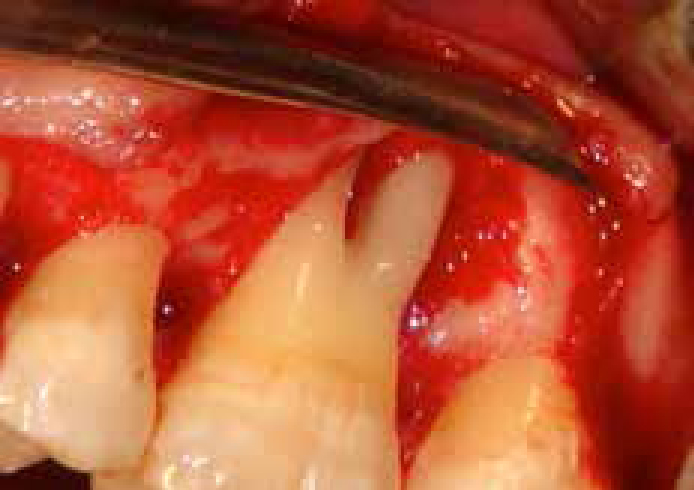
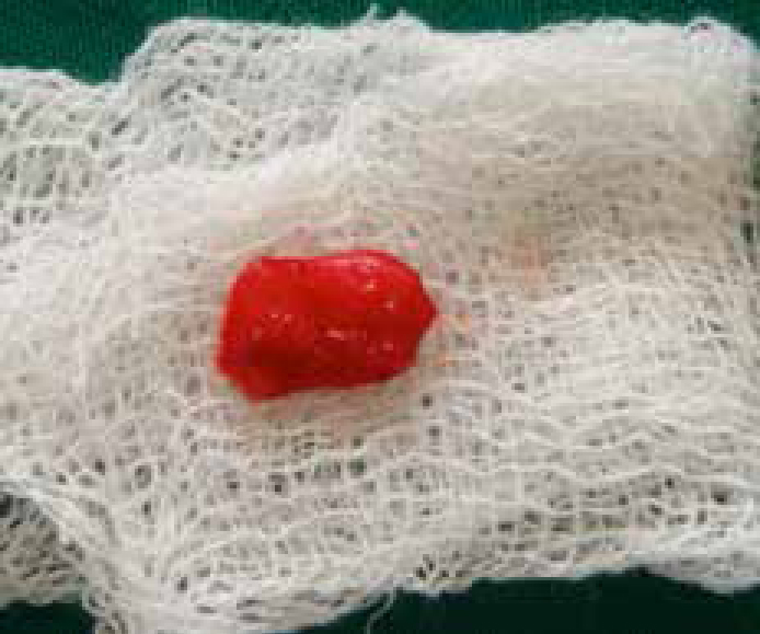
The surgical site was anaesthetized by the local administration of 2% lignocaine hydrochloride (LOX, Neon Laboratories Ltd, Mumbai, India) with 1:200000 adrenaline. A localized mucoperiosteal flap extending between UL4 and UL7 was raised buccally and palatally. The area was thoroughly debrided using area specific gracey curettes #9–10, #11–12 and #13–14 (Hu-Friedy Mfg Co, LLC) to reveal the bony defect at the furcation region of UL6 (Figure 3). After administration of local anaesthesia, the subepithelial connective tissue graft was harvested (Figure 4) and the epithelial door in the palatal donor site was sutured using an absorbable suture material (4-0 Vicryl, Ethicon, Inc, Johnson and Johnson, Somerville, NJ, USA). Next, bovine hydroxyapatite synthetic graft material (Bio – Oss, Geistlich Pharmaceuticals, Wolhausen, Switzerland) was carefully packed into the furcation region in UL6. Following this, the harvested subepithelial connective tissue graft was placed covering the bone graft to act as a membrane and was extended coronally to achieve root coverage simultaneously (Figure 5). The mucoperiosteal flap was sutured back in position in the buccal and palatal aspects using absorbable suture material (4-0 Vicryl, Ethicon Inc, Johnson and Johnson, Somerville, NJ, USA).
Figure 3. Mucoperiosteal flap raised and debrided revealing the bony defect at the furcation region of UL6.Figure 4. Subepithelial connective tissue graft harvested from the palate.Figure 5. Subepithelial connective tissue graft sutured in position at the recipient site in relation to UL6.
Post-operative instructions
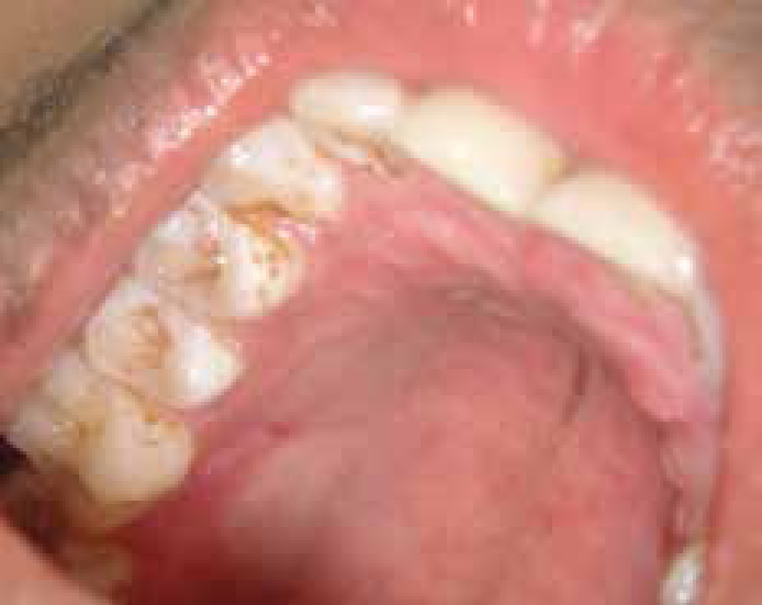
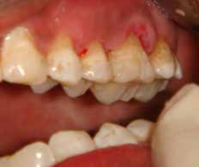
A prescription of 500 mg of amoxicillin three times a day for five days and 400 mg of metronidazole twice daily for five days was given. An analgesic, Ibuprofen, was also given; 400 mg three times a day for three days. The patient was asked to rinse with warm saltwater for the first two weeks to promote healing. Following this, the patient was instructed to use chlorhexidine gluconate mouthrinse 0.12% (Peridex, Zila Pharmaceuticals, Phoenix, AZ, USA) to facilitate plaque control.4 The patient was recalled and surgical sites were evaluated for wound healing at regular intervals. There was adequate root coverage and complete absence of pocketing and furcation defect in the treated site, with complete healing of the donor site in the palate (Figures 6 and 7). However, the recipient site showed marginal gingival tissue inflammation during the two-week post-operative check-up. The reason for this transient inflammation could be attributed to the periodontal pack placement for site protection which limited adequate oral hygiene practices in that region. The patient was advised not to use a toothbrush or any interdental cleaning aids during the first two weeks post-surgery, as the periodontal pack was in place, and he was advised to use mouthwash to rinse the debris at the surgical site. The subsequent recall visits showed significant wound healing, and one year following treatment the patient continued exhibiting adequate gingival recession coverage and a healthy periodontium (Figure 8). The patient continues to be on a strict home care regimen and regular recall visits to the dental practice.
Figure 6. Two weeks post-operative intra-oral view of the healed palate.Figure 7. Two weeks post-operative intra-oral view of healed recipient site in relation to UL6.Figure 8. One year post-operative intra-oral view of healed recipient site in relation to UL6.
Discussion
Periodontitis is a multifactorial, complex disease requiring therapy from various aspects. Periodontal therapy is primarily aimed at achieving a decrease in the microbial load and achieving periodontal attachment apparatus regeneration whenever feasible. Soft tissue replacement grafts have become a substantial element to increase tissue volume in plastic and aesthetic periodontal surgery and are commonly used to restore and/or improve mucogingival deformities and conditions around teeth or on edentulous ridges.5 Autogenous subepithelial connective tissue grafts are increasingly applied in aesthetic indications, like soft tissue thickening, recession treatment, ridge preservation, soft tissue ridge augmentation and papilla re-construction.6 Subepithelial connective tissue grafts have distinct advantages over free gingival autografts and other available synthetic graft options due to the rich vascular connective bed of this graft, as well as the natural colour-matching properties with the surrounding recipient tissues. The success of the subepithelial connective tissue graft is attributed to the double blood supply at the recipient site from the underlying connective tissue base and the overlying recipient flap.7 The donor site morbidity and patient discomfort is also reduced due to the suturing back of the epithelial door to its original pre-operative position. A study by Sedon et al concluded that subepithelial connective tissue grafting (SCTG) and guided tissue regeneration (GTR) techniques should be considered as the treatments of choice for root coverage. The results of this study demonstrated high and predictable root coverage of 98.9% and 92.3%, with the use of SCTG and GTR techniques, respectively.8 This case report demonstrated the successful use of subepithelial connective tissue graft in a dual role for achieving root coverage, as well as serving the purpose of a barrier membrane in guided tissue regeneration.
Conclusion
For the clinical performance of connective tissue graft harvesting and transplantation, a fundamental understanding of the anatomy at the donor sites and a sound knowledge of tissue integration and re-vascularization processes are required. Due to the distinct advantages of the subepithelial graft tissue, this tissue can be utilized with variant surgical techniques for achieving predictable and successful periodontal tissue regeneration.