Heyman SN, Babayof I. Emphysematous complications in dentistry, 1960–1993: an illustrative care and review of the literature. Quintessence Int. 1995; 26:535-543
McKenzie S, Rosenberg M. Iatrogenic subcutaneous emphysema of dental and surgical origin: a literature review. J Oral Maxillofac Surg. 2009; 67:1265-1268
Barkdull TJ. Pneumothorax during dental care. J Am Board Fam Pract. 2003; 16:165-169
Andsberg V, Axell T. Mediastinal emphysema as a complication of dental treatment. Odontol Res. 1072; 23:21-26
Chuong R, Boland TJ, Piper MA. Pneumomediastinum and subcutaneous emphysema associated with temporomandibular joint surgery. Oral Med Oral Surg Oral Pathol. 1992; 74:2-6
Snyder MD, Rosenberg ES. Subcutaneous emphysema during periodontal surgery: report of a case. J Periodontal. 1977; 48:790-791
Piecuch JF, West RA, Wash S. Spontaneous pneumomediastinum associated with orthognathic surgery. A case report. Oral Med Oral Surg Oral Pathol. 1979; 48:506-508
Lloyed RE. Surgical emphysema: a complication in endodontics. Br Dent J. 1975; 138:393-394
Carmichael F, Ward-Booth RP, Banks JM. Pneumomedistianum after facial trauma. Oral Med Oral Surg Oral Pathol. 1988; 66:540-542
Surgical emphysema: a rare complication of a simple surgical dental extraction without the use of an air-driven rotor Keegan Gowans Muneer Patel Khari Lewis Dental Update 2024 44:3, 707-709.
Authors
KeeganGowans
BDS, MJDF RCS(Eng), MSc
(Orthodontic Specialty Registrar), East Kent University Hospital Foundation Trust, William Harvey Hospital, Kennington Road, Willesborough, Ashford, Kent TN24 0LZ, UK
(Staff Grade Oral Surgeon), East Kent University Hospital Foundation Trust, William Harvey Hospital, Kennington Road, Willesborough, Ashford, Kent TN24 0LZ, UK
Surgical emphysema is a rare complication of dental extractions, often associated with the use of high-speed air rotors. This report describes a case of extensive surgical emphysema following a simple surgical extraction of a LL6 under local anaesthetic. There was no use of air-driven handpieces during the procedure. The patient developed extensive surgical emphysema bi-laterally in both cervical neck and facial planes. After prophylactic antibiotics with careful monitoring in a secondary care setting, the patient made a full unremarkable recovery.
CPD/Clinical Relevance: Simple extraction of teeth is a procedure carried out daily by most general dental practitioners. However, the risk of surgical emphysema without the use of high-speed air rotors or instruments using pressurized air/water is not well known or documented.
Article
This case report is one of cervicofacial surgical emphysema after a simple surgical extraction of a lower left six (LL6) under local anaesthetic. There was no use of a high-speed air rotor, and the most likely cause is an increase in intra-oral air pressure during a football training session two hours post-surgery. Our search of the literature yielded very few previously published reports of extensive emphysema without the use of equipment using pressurized air.
Surgical emphysema is a rare complication of many dental procedures, however, it can have serious and even life-threatening effects if not diagnosed and treated promptly. Potential spread of microbes such as staphloccocus, streptoccocal and anaerobes into tissue spaces is the cause of many complications. Most reported cases associated with dental procedures come from the use of air rotors or air syringes as reported by Heyman and Babayof1 and McKenzie and Rosenberg2 in systematic reviews. There have been few reported cases of surgical emphysema post-extraction when there has not been the use of air rotors during the removal.
Clinical findings
A 35-year-old male was referred to the accident and emergency department (A+E) at 04:00 with bilateral neck, left submandibular and left facial swelling. Twelve hours previously he had undergone a simple surgical extraction of the LL6 under local anaesthetic by a specialist oral surgeon in a general dental practice setting. Only an envelope flap was raised and a small amount of buccal bone removed using an electric motor handpiece. The extraction was described as unremarkable and the tooth removed without any complications.
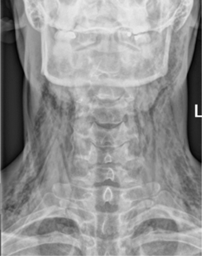
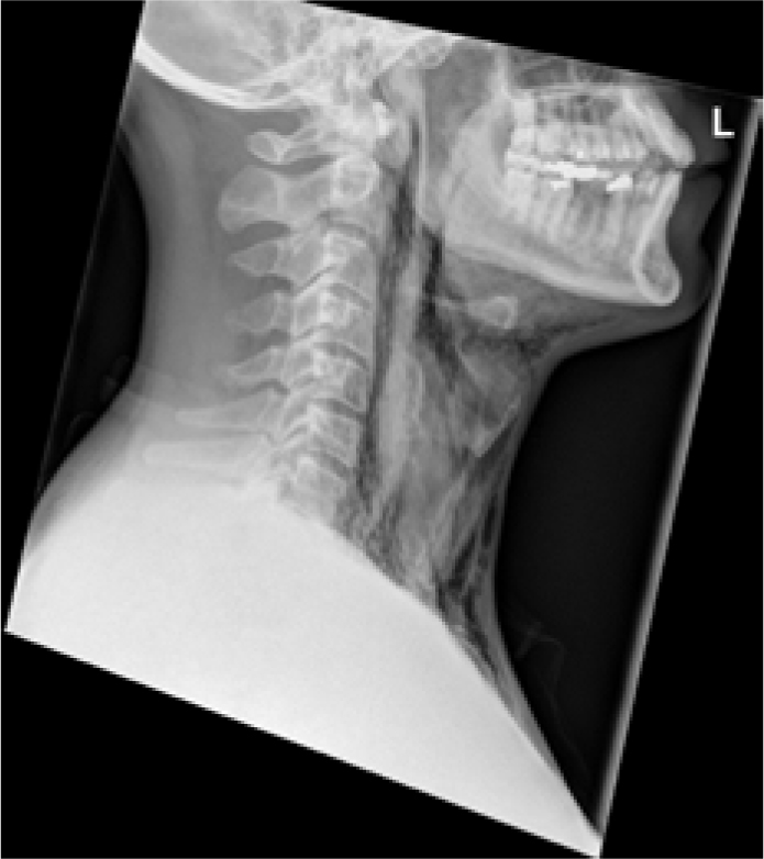
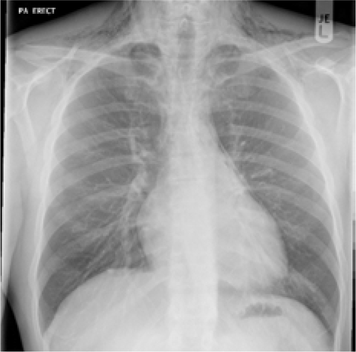
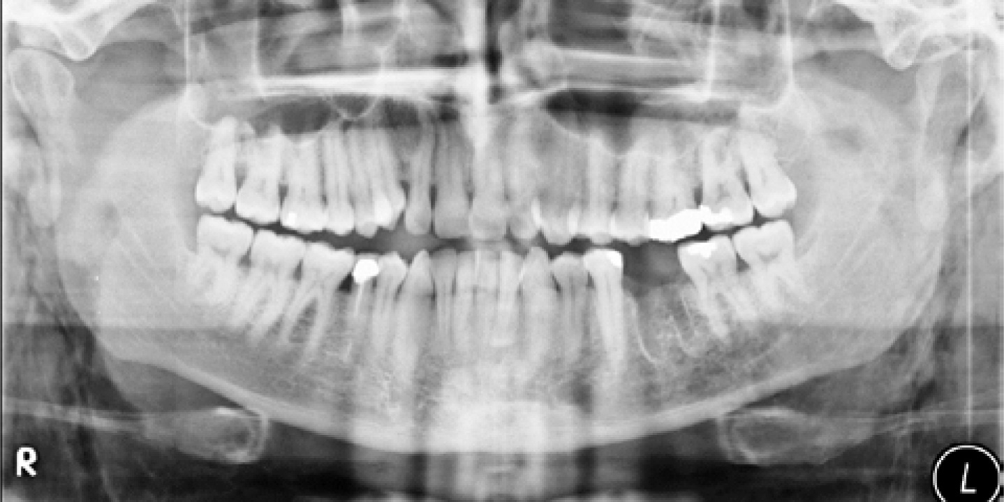
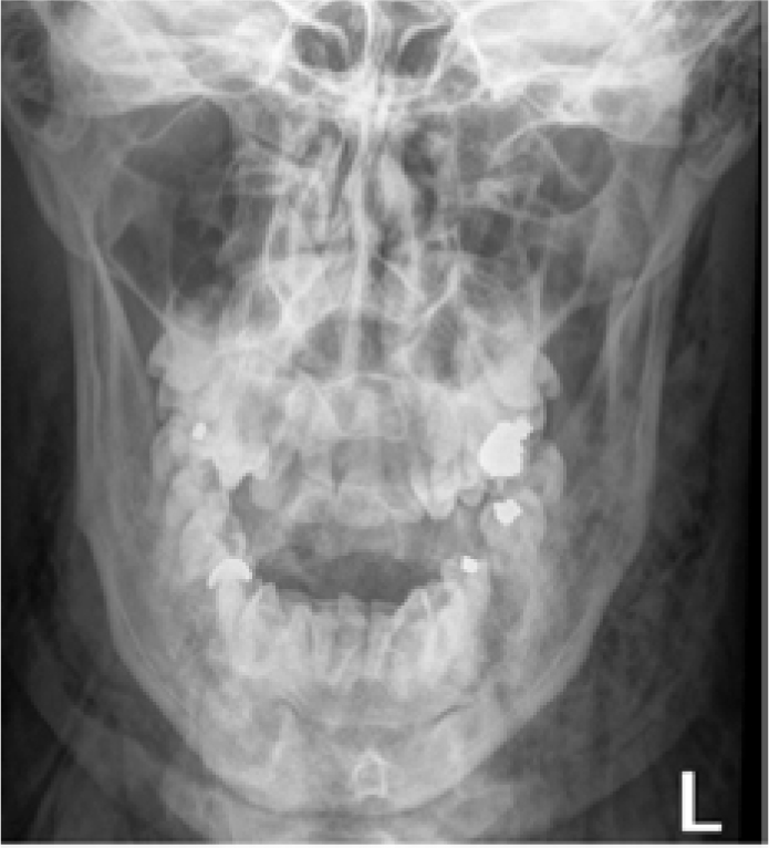
On presentation to A+E his pulse was 70/min, blood pressure 120/70 mm Hg, respiratory rate 14/min, temperature 36.9 °C and an oxygen saturation of 99%. On clinical examination there was palpable crepitus bilateral of his cervical neck, left submandibular region and left face extending to the left temple region. Radiographic examination of his chest, neck and mandible showed that there was extensive surgical emphysema around the anterior neck and mandible. Radiographic examination of the neck showed air within the cervical tissue planes (Figures 1 and 2). A chest radiograph showed no evidence of mediastinal emphysema (Figure 3). It was evident from both the OPG (Figure 4) and the PA mandible (Figure 5) that there was bilateral spread of air through the facial tissue planes.
Figure 1. Soft tissue neck view showing extensive bilateral neck emphysema.Figure 2. Soft tissue lateral neck view.Figure 3. Chest radiograph showing surgical emphysema.Figure 4. OPG showing the extraction socket of the LL6. Septal bone is still present showing very little bone removal during the extraction.Figure 5. PA mandible showing extensive facial surgical emphysema.
Treatment
The patient refused admission to hospital, therefore treatment consisted of oral antibiotics of Co-amoxiclav 625 mg (500/125) TDS for 7 days and Metronidazole 400 mg TDS for 5 days. A review was undertaken at 1 day, 3 days and 2 weeks.
On day 1 there was little change to the swelling or the patient's symptoms of neck stiffness and pain. However, by day 3 there was very little residual swelling. By day 14, the crepitus had dissipated and a full recovery was made without further complications.
Discussion
In this case, there was no use of an air rotor or 3-in-1 syringe which could force air into the tissue spaces. The tooth was extracted by a specialist oral surgeon in a primary care setting and delivered with forceps. After taking a detailed history of events from the patient, the only potential cause for the surgical emphysema was that he attended a football training session, where he is the coach, 2 hours post-operatively. The use of a whistle and/or cardiovascular exercise can lead to increased pressure within the oral cavity on exertion, forcing air into tissue spaces through the extraction site. The fact that the patient did not have any swelling immediately post-operatively, and only reported swelling after the football training session, is further evidence of self-induced emphysema.
The spread of large amounts of air into deep neck spaces may sometimes cause serious complications, including cardiac and respiratory failure from pneumopericardium, pneumomediastinum, or airway compromise due to the accumulation of air in the retropharyngeal space.3 It is therefore essential that a swift diagnosis is made and relevant investigations carried out.
Use of air rotors during surgical extraction of teeth is the most commonly reported cause of surgical emphysema post-dental extraction. However, other dental procedures, including restorative, periodontal, endodontic, orthognathic surgery and facial trauma have all been implicated.4,5,6,7,8,9 Heyman and Babayof1 looked at 74 cases of emphysema following dental procedures between 1960 and 1993. They found that air rotors or air syringes were implicated in 71% of reported cases. A more recent systematic review carried out by McKenzie and Rosenberg2 looked at cases between 1993 and 2008, when there were 32 reported cases of surgical emphysema. Of these cases, 50% were linked to air rotors. However, other causes were reported as a CO2 laser, a NO2 cryomachine, an air abrasive system and an endotracheal intubation/ventilation.
As described in many case reports, treatment is largely symptomatic relief and administration of antimicrobials, ideally administered intravenously, with hospital admission to enable regular monitoring of the patient's vital signs. An antibiotic is needed which covers for normal oral flora. In this case, the patient refused admission to hospital and was therefore given oral Co-Amoxiclav and a follow-up appointment the following day for monitoring.
Conclusion
This case highlights the need for all patients to be given adequate post-operative instructions, including minimal physical activity post-surgery to minimize the risk of this sometimes life-threatening complication.