The glossary of prosthodontic terms. J Prosthet Dent. 1994; 71:41-112
Jacobson TE, Krol AJ A contemporary review of the factors involved in complete denture retention, stability, and support. Part I: retention. J Prosthet Dent. 1983; 49:5-15
Jacobson TE, Krol AJ A contemporary review of the factors involved in complete dentures. Part II: stability. J Prosthet Dent. 1983; 49:165-172
Scott BJ, Hunter RV Creating complete dentures that are stable in function. Dent Update. 2008; 35:259-267
Kelly E Changes caused by a mandibular removable partial denture opposing a maxillary complete denture. J Prosthet Dent. 1972; 27:140-150
Witter DJ, van Palenstein Helderman WH, Creugers NH, Kayser AF The shortened dental arch concept and its implications for oral health care. Community Dent Oral Epidemiol. 1999; 27:249-258
McCord JF, Grant AA Registration: stage III – selection of teeth. Br Dent J. 2000; 188:660-666
Lynch CD, Allen PF Management of the flabby ridge: using contemporary materials to solve an old problem. Br Dent J. 2006; 200:258-261
Carlsson GE Facts and fallacies: an evidence base for complete dentures. Dent Update. 2006; 33:134-142
For many years, it has been considered necessary to restore posterior support with a bilateral free-end saddle when a full upper denture is opposed by a shortened dental arch. It was thought that this would provide occlusal stability and prevent anterior bite collapse and temporomandibular dysfunction. As free-end saddle partial dentures are often poorly tolerated by patients, this case study tests whether a retentive full-upper denture occluding with a shortened dental arch offers enough to fulfil a patient's needs.
CPD/Clinical Relevance: An increasingly elderly population will present dental practitioners with more partially dentate patients requiring treatment.
Article
It has been traditionally accepted that a full upper denture requires good retention and stability to be of functional use to a patient. Authors have separated the principles of retention, resistance to displacement along the path of insertion, and stability, the resistance of a denture to displacement by functional forces.1,2,3 These forces may be forces acting to displace the denture in many different directions.4
Historically, an edentulous maxillary arch opposed by a shortened mandibular arch would have been treated with a complete upper denture and a bilateral free-end saddle removable partial denture (RPD).5 Since then, significant research has led to the shortened dental arch (SDA) concept which states that 10 occluding pairs of teeth are sufficient for masticatory function.6 With this in mind, is the looseness of a maxillary complete denture occluding with a SDA attributable to a lack of posterior support or can it be corrected by a more accurate reproduction of the denture-bearing area?
Clinical report
A 68-year-old female with an edentulous maxilla and a shortened mandibular dental arch presented complaining of a loose full upper denture. She had a 35-year history of wearing upper dentures, the current prosthesis being five years old. She thought she had about nine in all and had been using a fixative to secure her dentures for as long as she could remember. The current set were the most comfortable she had worn and were made using a copy technique from her previous denture. There was a mention of sensitivity to zinc oxide impression paste recorded in the patient's notes when this denture had been made. The upper left central incisor had been re-attached following a fracture. She had never been provided with a lower partial denture and considered that she managed perfectly well providing she had fixed her upper denture in place. Her medical history revealed that she was fit and well. She used levothyroxine for hypothyroidism and had a latex allergy.
Her face appeared symmetrical. On intra-oral examination, it was noted that the patient had a traumatic ulcer on the buccal mucosa where she had bitten her cheek. The buccal cusps of the upper artificial molars had been adjusted to alleviate this in the past. The artificial premolars and anterior teeth appeared evenly worn. The post dam was well short of the vibrating line. The occlusion was balanced with slight displacement in lateral excursions but the patient was able to secure the denture with the posterior of her tongue on incisal contact. She had fixative adhered to the fitting surface to keep the denture in place.
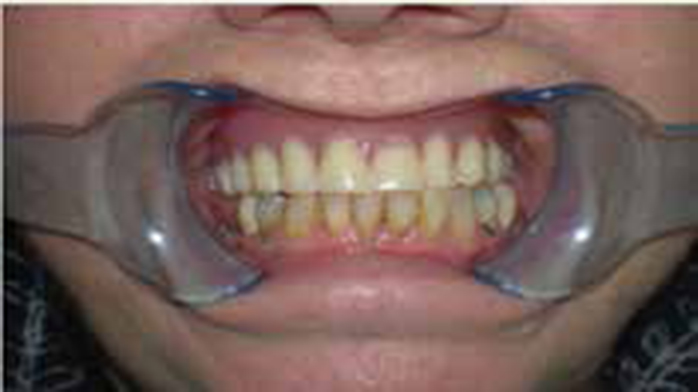
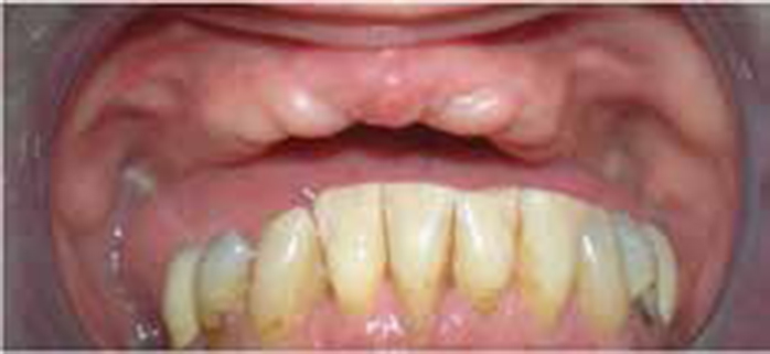
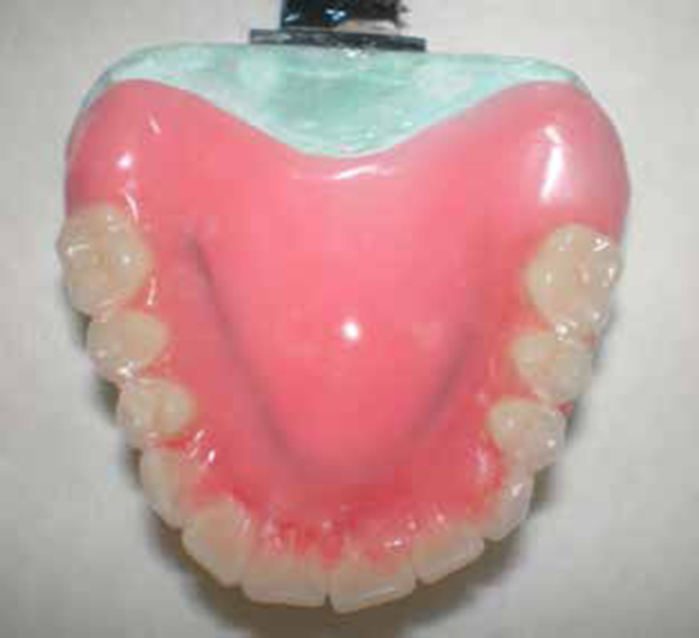
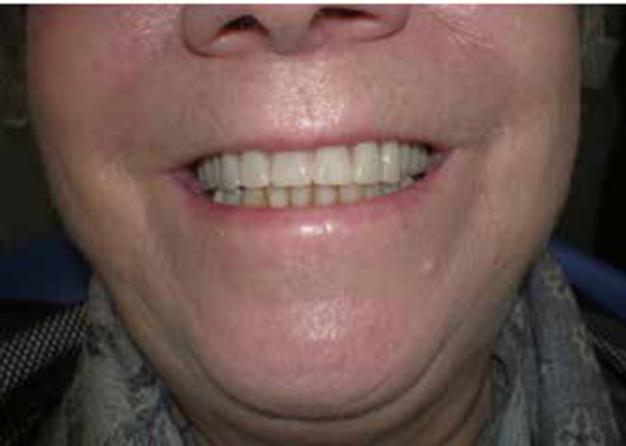
She had 10 sound, remaining lower teeth (LL5–LR5) and both second premolars had porcelain-bonded crowns. Her periodontal condition showed some bleeding on probing with pocket depths not exceeding 2 mm. She had some supragingival calculus on the lingual surfaces of her lower incisors. The edentulous upper ridge showed some signs of alveolar bone loss and replacement with fibrous tissue anteriorly and there was some papillary hyperplasia of the hard palate. The maxillary tuberosities did not appear to be particularly overgrown. Following consent from the patient, photographs were taken for inclusion in a case study (Figures 1 and 2).
Figure 1 Old denture in situ.Figure 2. Denture-bearing area with flabby anterior ridge.
After discussion with the patient considering the different treatment options, it was decided to accept her lower arch and provide her with a new full upper denture, hopefully with improved retention and stability. The evenly worn occlusion would suggest chopping vertical mandibular movements during mastication.7 She was asked whether she had any old photographs of her showing her natural teeth, which she agreed to provide, but she was happy with the uniformity of her current denture's appearance. The detail of the anterior flabby ridge would need to be recorded accurately and the posterior border seal would need to make better use of the available area. Green-stick compound was added to the posterior border of her old denture to extend the post dam temporarily. She was able to tolerate this and an improvement in retention was noted immediately. The green-stick was removed from the denture before the patient left the surgery. Extending the position of the post dam on the new denture would increase retention. It was also considered unnecessary to add second maxillary molars, as these would be functionless and possibly contribute to her buccal cheek trauma.
A preliminary impression of the mandibular arch and the maxillary denture-bearing area was made with a low viscosity hydrocolloid (DE Healthcare Alginate). The area of displaceable (flabby) tissue was indicated on the impression for the technician to incorporate a spacer into the upper special tray in this area.
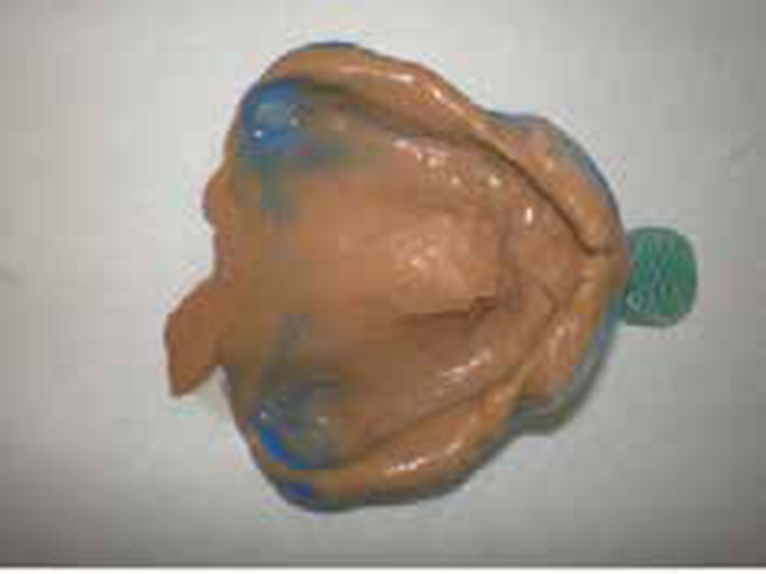
The secondary impression was taken as a two-stage technique with silicone impression material, as described by Lynch and Allen.8 Heavy-bodied material (Henry Schein, UK) was used, first paying careful attention to border mould the sulcus area. The heavy-bodied silicone was then removed from the relieved part of the special tray over the displaceable area with a scalpel when fully set. Then a second stage impression was taken with mucostatic light-bodied silicone (Virtual, Ivoclar Vivadent, Liechtenstein) to limit the displacement of the flabby ridge (Figure 3). The intended position of the post dam was recorded for the technician. Carlsson's evidence-based review of complete dentures describes a series of 20-year randomized controlled trials that refuted the use of sophisticated instruments in denture registration. Edentulous patients were allocated to two treatment groups, one using a complex technique involving hinge-axis location for a facebow transfer to the articulator and the other without facebow, only an arbitrary mounting. No significant difference was found.9 He went on to say that facebows have been abandoned long ago in Scandinavia for removable and fixed prosthodontic work. The wear pattern of the old denture in this case would suggest a lack of ruminatory masticatory movements anyway.7 Jaw registration was recorded with wax rims and sent to the technician for an arbitrary mounting.
Figure 3. Mucostatic impression.
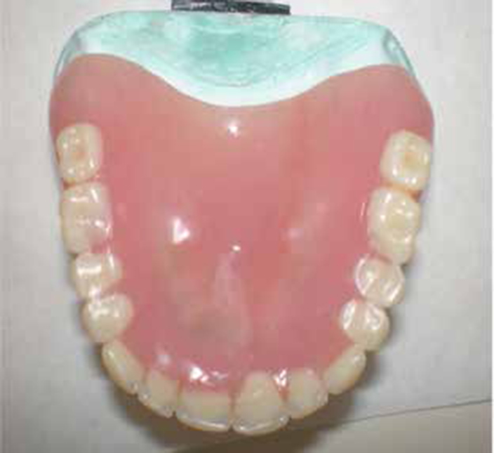
The try-in stage can be seen compared with the old denture (Figures 4 and 5) with the new post dam position giving considerable additional posterior coverage. Efforts were made to rotate the artificial lateral incisors to re-create the patient's smile from one of the old photographs she supplied, but she was unhappy with a deviation from the perfect uniform set-up to which she had become accustomed. The dentures were delivered and, after minor adjustments with denture indicator spray (Hydent), the patient was very satisfied with the fit (Figure 6). Lateral and protrusive excursions were checked and adjusted for balancing with horseshoe articulating paper. At one-week review, the patient reported that she had not required fixative even to eat a meal. She was still experiencing a little soreness around the flange in the tuberosities and she mentioned biting her cheeks. The buccal cusps of the artificial first molars were adjusted. She cancelled her second review a fortnight later as she stated that she was completely satisfied.
Figure 4. Old denture on cast.Figure 5. Try-in stage.Figure 6. Denture at fit stage.
Discussion
The basic principles for upper complete denture construction ring true in this case study. The patient's complaint of a loose denture was treated by provision of a denture made by impressions recording good surface detail extending over the whole of the denture-bearing area. The key messages to be taken from this case are as follows.
Carefully assess the extensions
An adequate peripheral seal is crucial with the post dam lying just short of the vibrating line with extension around the tuberosities.
Appropriate impression technique is required for a flabby ridge
The previous denture was made using a copy technique and it would not have been possible to control the movement of the fibrous tissue with a closed mouth impression technique used in the copying process. For this reason, the tissues in this area need to be recorded using a mucostatic impression. The anterior ‘flabby’ area of the maxillary edentulous ridge will tend to dislodge the denture when not loaded occlusally as it recoils from compression.10
Occlusion is critical
The accurate impression technique and well extended peripheral seal will be broken if the occlusion is ignored. Displacing forces are reduced by matching the maxillary artificial teeth and maxillary occlusal plane to mandibular movement. There should be articulating contacts balanced across multiple teeth on the denture during excursive movements.11
Interestingly, despite evidence of improved retention in this case being supported by the temporary green-stick post dam, Floystrand et al, reported insignificant reduction of retention of maxillary dentures following extensive reduction of the palatal coverage. They found that the border could form a U-shape 10 mm from the dental arch and still be as retentive.9
Conclusion
A denture solution has been achieved in this case by listening to the patient's complaints, looking at the shortcomings of the patient's previous denture and using a technique for accurately measuring the denture-bearing area of the maxilla. The patient's stable, opposing SDA and perhaps her high tolerance level from unstable previous dentures has enabled a simplified registration stage too.