Marx RE Pamidronate (Aredia) and zoledronate (Zometa) induced avascular necrosis of the jaws: a growing epidemic. J Oral Maxillofac Surg. 2003; 61:1115-1117
Fliefel R, Tröltzsch M, Kühnisch J, Ehrenfeld M, Otto S Treatment strategies and outcomes of Bisphosphonates related osteonecrosis of the jaw (BRONJ) with characterisation of patients: a systematic review. Int J Oral Maxillofac Surg. 2015; 44:568-585
Ruggiero SL, Dodson TB, Fantasia J American Association of Oral and Maxillofacial Surgeons position paper on medication related osteonecrosis of the jaws – 2014 an update. J Oral Maxillofac Surg. 2014; 72:1938-1956
Migliorati CA, Siegel MA, Elting LS Bisphosphonate-associated osteonecrosis: a long-term complication of bisphosphonate treatment. Lancet Oncol. 2006; 7:508-514
Reid IR Osteonecrosis of the jaw: who gets it, and why?. Bone. 2009; 44:4-10
Hansen T, Kirkpatrick CJ, Walter C, Kunkel M Increased numbers of osteoclasts expressing cysteine proteinase cathepsin K in patients with infected osteoradionecrosis and bisphosphonate-associated osteonecrosis – a paradoxical observation?. Virchows Archiv. 2006; 449:448-454
Sedghizadeh PP, Kumar SKS, Gorur A, Schaudinn C, Shuler CF, Costerton JW Identification of microbial biofilms in osteonecrosis of the jaws secondary to bisphosphonate therapy. J Oral Maxillofac Surg. 2008; 66:767-775
: Dental Clinical Guidelines; 2011
Zaiirowski J Comment on the American Association of Oral and Maxillofacial Surgeons statement on bisphosphonates. J Oral Maxillofac Surg. 2007; 65:1440-1441
Ruggiero SL, Mehrotra B, Rosenberg TJ, Engroff SL Osteonecrosis of the jaws associated with the use of bisphosphonates: a review of 63 cases. J Oral Maxillofac Surg. 2004; 62
Hillner BE, Ingle JN, Chlebowski RT American Society of Clinical Oncology 2003 update on the role of bisphosphonates and bone health issues in women with breast cancer. J Clin Oncol. 2003; 21:4042-4057
Katz J, Gong Y, Salmasinia D Genetic polymorphisms and other risk factors associated with bisphosphonate induced osteonecrosis of the jaw. Int J Oral Maxillofac Surg. 2011; 40:605-611
Nicoletti P, Cartsos VM, Palaska PK, Shen Y, Floratos A, Zavras AI Genome wide pharmacogenetics of bisphosphonate-induced osteonecrosis of the jaw: the role of RBMS3. Oncologist. 2012; 17:279-281
Marini F, Tonelli P, Cavalli L Pharmacogenetics of bisphosphonate-associated osteonecrosis of the jaw. Front Biosci (Elite Ed). 2011; 3:364-370
AHFS Consumer Medication Information [Internet]. 2015. (Accessed 31/05/15)
Allegra A, Oteri G, Alonci A Association of osteonecrosis of the jaws and POEMS syndrome in a patient assuming rituximab. J Craniomaxillofac Surg. 2014; 42:279-282
Weighert KL, Lewgoy J, Mazzoleni DS, Franco FR, Enriconi L, Sasso JH Rituximab and osteonecrosis of the jaws: case study. Oral Surg Oral Med Oral Pathol Oral Radiol. 2014; 117:188-189
Joint Formulary Committee. British National Formulary (Internet). (Accessed on 19/10/15)
Guarneri V, Miles D, Robert N Bevacizumab and osteonecrosis of the jaw: incidence and association with bisphosphonate therapy in three large prospective trials in advanced breast cancer. Breast Cancer Res Treat. 2010; 122:181-188
Hasewgawa Y, Kawabe M, Kimura H, Kurita K, Fukuta J, Urade M Influence of dentures in the initial occurrence site on the prognosis of Bisphosphonate-related osteonecrosis of the jaws: a retrospective study. Oral Surg Oral Med Oral Pathol Oral Radiol. 2012; 114:318-324
Carlson ER, Basile JD The role of surgical resection in the management of bisphosphonate-related osteonecrosis of the jaws. J Oral Maxillofac Surg. 2009; 67:85-95
Rupel K, Ottaviani G, Gobbo M A systematic review of therapeutical approaches in Bisphosphonates-related osteonecrosis of the jaw (BRONJ). Oral Oncol. 2014; 50:1049-1057
Kyrgidis A, Vahtsevanos K, Koloutsos G Bisphosphonate-related osteonecrosis of the jaws: a case-control study of risk factors in breast cancer patients. J Clin Oncol. 2008; 26:4634-4638
Göllner M, Holst S, Fenner M, Schmitt J Prosthodontic treatment of a patient with Bisphosphonate-induced osteonecrosis of the jaw using a removable dental prosthesis with a heat-polymerized resilient liner: a clinical report. J Prosthetic Dent. 2010; 103:196-201
Conservative prosthetic rehabilitation of medication-related osteonecrosis of the jaw (MRONJ) Alexandra Johanna Leven Antony J Preston Dental Update 2024 43:10, 707-709.
Authors
Alexandra JohannaLeven
BDS, MFDS RCSEd
Specialty Registrar in Restorative Dentistry, Liverpool University Dental Hospital, Pembroke Place, Liverpool L3 5PS, UK (a.leven@nhs.net)
Osteonecrosis of the jaw associated with bisphosphonates and other medications is a growing problem facing dentists. It can have a significant and debilitating impact upon patients. Various treatment options ranging from surgical intervention to management with antibiotics and analgesics have been proposed. This article presents one method of conservative treatment and prosthetic rehabilitation in a patient with ongoing BRONJ of the maxilla unsuitable for surgical management.
CPD/Clinical Relevance: Dentists need to be able to identify patients who are at risk of developing BRONJ and have an awareness of the appropriate management as well as potential oral rehabilitation options for these patients.
Article
First reported by Marx in 2003,1 bisphosphonate-related osteonecrosis of the jaw (BRONJ) continues to be an increasing problem facing the dental and medical profession.
The anti-resorptive and anti-angiogenic properties of bisphosphonates (BPs) give them a role in the management of various skeletal conditions, such as osteoporosis, osteopenia and Paget's disease, as well as in the treatment of multiple myeloma, prostate, lung and breast cancer.2
The side-effects of BPs on the alveolar bone is well documented, however, the aetiology is not fully understood. Other bones are seemingly unaffected – this may be explained by higher bone turnover rate of the jaws compared with other bones and bacterial ingress from the teeth and periodontium.3,4 Various theories have been proposed to explain the pathophysiology of BRONJ. These are discussed in more detail in other publications,2,3,4,5 however, two proposed theories are broadly as follows:
The ‘inside out theory’ where localized bone micro-trauma is in combination with reduced osteoclast activity; and
Bone remodelling results in necrosis of bone and soft tissue.2,4
However, several studies now dispute reduced bone turnover as a factor.5,6 Conversely, the ‘outside in theory’ suggests bacteria from the oral cavity colonize the exposed bone following a traumatic event, such as tooth extraction or denture trauma, which leads to necrosis of the underlying bone. A variety of bacterium species have been isolated but actinomyces has been a common finding in osteonecrosis of the jaw (ONJ).5,7 However, BRONJ is more likely to be a multifactorial condition relying on the presence of multiple host factors before it occurs.2 There are numerous risk factors that increase the potential for BRONJ and these must be considered when planning treatment (Table 1).
Higher affinity bisphosphonate, ie zolendronate
Higher potency bisphosphonate, ie zolendronate
Prolonged use of bisphosphonates of 3 years or more8
Invasive dental treatment or oral soft tissue trauma3
*Osteonecrosis affects females more than males (2:1).2 However, that may be due in greater part to the fact that osteoporosis is more common in females, meaning bisphosphonate use is higher in the female population rather than these patients being more susceptible to osteonecrosis.2
Bisphosphonates are not the only drugs to cause ONJ and, therefore, it has been proposed that medication-related osteonecrosis of the jaws (MRONJ) would be a more appropriate term.3 Denosumab (Prolia®, Amgen, Thousand Oaks, CA, USA), a RANK ligand inhibitor used in the treatment of osteoporosis, can result in the same oral presentation.3,15 ONJ has also been reported in patients taking the following medications: tyrosine kinase inhibitors, eg Sunitinib (Sutent®, Pfizer, New York City, NY, USA); monoclonal antibodies targeting vascular endothelial growth factor, eg Rituximab (Rituxan®, Biogen Idec Inc, Weston, MA, United States) and Bevacizumab (Avastin®, Genetech, San Francisco, CA, United States), used to treat various cancers.3,16,17,18,19 Dentists must be aware of these additional medications and screen patients appropriately prior to extractions and other similar surgical procedures.
A range of treatment options, from major surgical intervention to more conservative management with antibiotics and analgesics, have been proposed. However, there is as yet no consensus on what the gold standard treatment should be. The American Association of Oral and Maxillofacial Surgeons (AAOMS) staging criteria and treatment for MRONJ gives a useful guide as to the severity of the condition and how it might be managed (Table 2).
Stage
Signs/Symptoms
Treatment
At Risk
No exposed bone but taking BPT
Patient education
Stage 0
No exposed bone but nonspecific signs/symptoms
Analgesics/antibiotics
Stage 1
Asymptomatic exposed bone
Antiseptic mouthwash Monitor 3/12
Stage 2
Symptomatic exposed bone, associated infection
AntibioticsAntiseptic mouthwashSuperficial debridement to ease soft tissue irritation
Stage 3
Symptomatic exposed bone and infection. Pathologic fracture/extra-oral fistula/osteolysis to inferior border of mandible
Antiseptic mouthwash. Antibiotics, analgesia.Surgical debridement or resection for long-term palliative care
In the current literature there is increased support for surgical intervention to remove bone sequestra, facilitate debridement and promote healing, particularly in symptomatic patients.3,20,21 A recent systematic review found that extensive surgical intervention or laser surgery resulted in the highest healing rates (80–100%).22 However, for some patients surgical treatment is not an option owing to other medical issues and MRONJ may become a chronic longstanding problem with limited resolution. Despite this, patients may seek prosthetic rehabilitation for aesthetic and functional reasons. This case report highlights one method of conservative prosthetic rehabilitation in a patient unsuitable for surgical intervention.
Case report
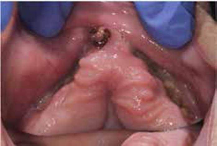
An 85-year-old female edentulous patient was referred by her GDP with exposed bone in the anterior region of the maxilla. She was treated jointly by the Oral Surgery and Restorative Dentistry departments. The patient experienced no pain but complained of a foul smelling/tasting discharge. On examination, two areas of exposed necrotic bone were noted bilaterally in the anterior maxilla; the areas were separated by a thin area of keratinized mucosa and a retained UR1 root (Figure 1). There was suppuration present but no loose bone fragments.
Figure 1. Bilateral BRONJ of anterior maxilla at initial presentation.
The patient was a wheelchair-user and had a complex medical history including: osteoporosis, under active thyroid, atrial fibrillation, asthma, hypertension, acid reflux and her current medication included bisoprolol, co-codamol, furosemide, hypromellose, levothyroxine, mometasone furoate, omeprazole, pregabalin, quinine sulphate, salbutamol and warfarin. The oral bisphosphonate, ibandronate sodium, was prescribed for more than 10 years for the management of the patient's osteoporosis. This was discontinued in December 2013.
Prior to referral, the patient had had a new set of complete dentures made. It was suspected that the upper denture had caused soft tissue trauma resulting in the exposed bone. In light of the patient's existing medical problems, invasive surgical treatment was deemed unsuitable. Instead, a conservative medical approach was employed. This involved chlorhexidine mouthrinses and prescription of long-term doxycycline and removal of loose bone fragments. This resolved the infection and discharge but initially no reduction in the surface area of exposed bone was observed. Despite this limited improvement, having been edentulous for over a year, the patient requested to have a further set of dentures made to improve aesthetics and function.
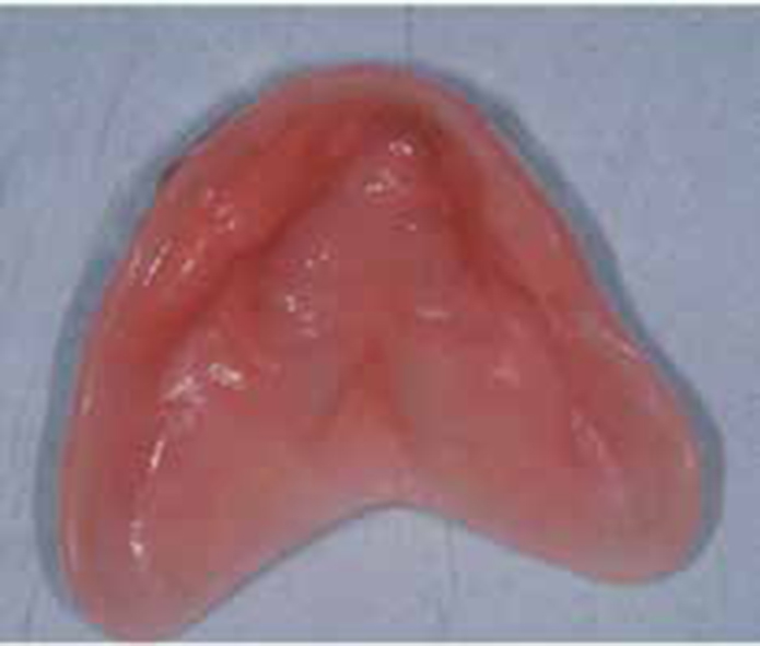
New acrylic dentures were constructed in a conventional manner. Primary alginate stock tray impressions were followed by medium-bodied silicone impressions in closely adapted special trays. Particular attention was paid to border-moulding with thermoplastic reline material (GC ISO Functional Sticks®). Registration was carried out using wax rims on heat-cured acrylic bases; this allowed the rims to be more stable and also provided insight as to the retention of the dentures at an early stage. For the try-in stage, a partial permanent soft reline material (Molloplast®) was placed around the areas of MRONJ to provide cushioning in these areas. However, this was taken a step further and a full, permanent, soft lining placed for the fit appointment (Figure 2). This not only improved retention further but, by providing a full soft lining, this limited any trauma to the whole of the denture-bearing area of the palate.
Figure 2. Permanent soft lining material on maxillary denture.
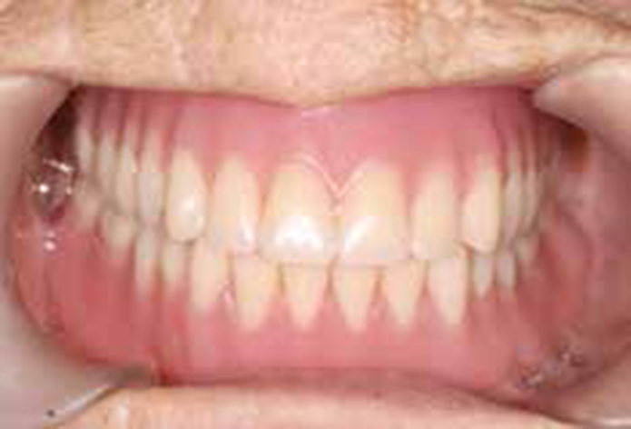
Given the amount of tissue necrosis, it was unclear what level of retention would be achievable with a denture; however, the result proved to be excellent. The patient and her family were very pleased with the result (Figure 3).
Figure 3. Finished complete set of acrylic dentures.
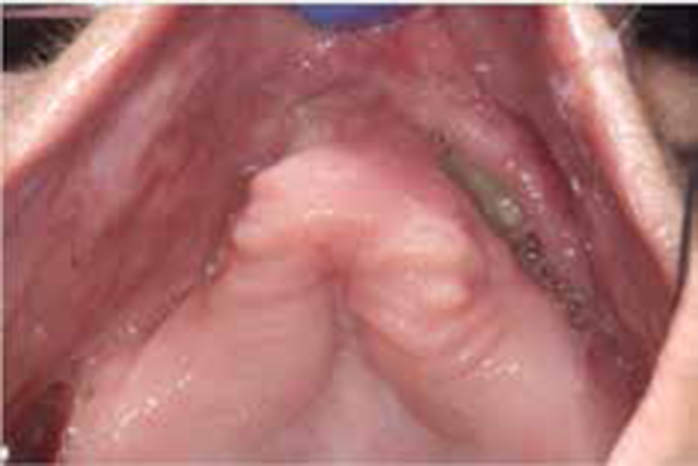
When the patient was reviewed 3 months later, she had lost approximately a 2 cm2 section of necrotic bone from the right maxilla and also the retained root. As a result, the upper denture had now become unstable. The denture was relined with further permanent soft lining in the laboratory. Again, this provided the patient with good retention and stability of the prosthesis. At a further 3 months follow-up appointment, the right maxillary region where the bone loss had occurred was noted to have healed significantly and there was now mucosal coverage in this area (Figure 4).
Figure 4. Some healing of right anterior maxilla following loss of bone sequestrum.
Given the progressive nature of this patient's MRONJ, it was reiterated that the denture may only be suitable for social use rather than daily function. Should the MRONJ progress, the prosthesis will have to be adapted further. However, for the time being the MRONJ has significantly improved following the loss of bone sequestrum in the area and so conservative treatment in this instance appears to have been effective.
Discussion
Although complete resolution of the patient's condition has not yet been achieved, this case serves to highlight a method of conservatively rehabilitating patients with MRONJ who are unsuitable for surgical intervention. Surgical intervention in a medically fitter patient may provide improved healing in areas of MRONJ but this should be assessed on a case-by-case basis. As has been demonstrated with this case, patients must be kept under close review to ease any traumatic areas of the denture and modify the denture base appropriately.
The link between dentures and the development of MRONJ is considered to be significant, with the mandibular molar region most frequently affected.20,23 It has been shown that denture wearers have a shorter duration to the onset of MRONJ compared to non-denture wearers.20 However, conversely, it is reported that, with good oral hygiene, dentures may in fact reduce recurrence of MRONJ post-treatment,20,24 possibly as, once the prosthesis is relieved or relined, it then protects the mucosa from external trauma.
Conclusion
Further long-term research is required to ascertain the gold standard management of medication-related osteonecrosis of the jaw. Until this can be established, each patient must be assessed on an individual basis and kept under close review. This paper has demonstrated one method of managing a case of MRONJ, however, this would clearly not be suitable for every patient. This may be a useful treatment for short-term rehabilitation while healing of a site of exposed bone is still ongoing.
Dentists must remain vigilant for signs of osteonecrosis, in not only patients prescribed bisphosphonates, but also RANK ligand inhibitors, tyrosine kinase inhibitors and monoclonal antibodies. Patients must be well informed so that they understand their role in maintaining good oral hygiene and seeking help from the dentist should they develop any signs or symptoms or their prosthesis causes trauma.