Secord PF, Backman CW Malocclusion and psychological factors. J Am Dent Assoc. 1959; 59:931-938
Nattrass C, Sandy JR Adult orthodontics – a review. Br J Orthod. 1995; 22:(4)331-337
Matthew SY, Chia F, Naini B, Gill DS The aetiology, diagnosis and management of mandibular asymmetry. Ortho Update. 2008; 1:44-52
Murphey MD, Choi JJ, Kransdorf MJ, Fleming DJ, Gannon HF Imaging of osteochondroma: variants and complications with radiologic-pathologic correlation. Radiographics. 2000; 20:1407-1434
Karasu HA, Ortakoglu K, Okcu KM, Gunhan O Osteochondroma of the mandibular condyle: report of a case and review of the literature. Mil Med. 2005; 170:(9)797-801
Ribas Mde O, Martins WD, De Sousa MH, Zanferrari FL, Lanzoni T Osteochondroma of the mandibular condyle: literature review and report of a case. J Contemp Dent Pract. 2007; 8:52-59
Kermer Ch, Rasse M, Undt G, Lang S Cartilaginous exostoses of the mandible. Int J Oral Maxillofac Surg. 1996; 25
Vezeau PJ, Fridrich KL, Vincent SD Osteochondroma of the mandibular condyle: literature review and report of two atypical cases. J Oral Maxillofac Surg. 1995; 53:954-963
Cimino R, Steenks MH, Michelotti A, Farella M, PierFrancesco N Mandibular condyle osteochondroma: review of the literature and report of a misdiagnosed case. J Orofac Pain. 2003; 17:254-261
Wolford LM, Mebra P, Franco P Use of conservative condylectomy for treatment of osteochondroma of the mandibular condyle. J Oral Maxillofac Surg. 2002; 60:262-268
Gaines RE, Lee MB, Crocker DJ Osteochondroma of the mandibular condyle; case report and review of the literature. J Oral Maxillofac Surg. 1992; 50
Zhang J, Wang H, Li X, Li W, Wu H, Miao J, Yuan X Osteochondromas of the mandibular condyle: variance in radiographic appearance on panoramic radiographs. Dentomaxillofac Radiol. 2008; 37:154-160
Karasu HA, Ortakoglu K, Okcu KM, Gunhan O Osteochondroma of the mandibular condyle: report of a case and review of the literature. Mil Med. 2005; 170:(9)797-801
Herbosa EG, Rotskoff KS Condylar osteochondroma manifesting as class III skeletal dysplasia: diagnosis and surgical approach. Am J Orthod Dentofacial Orthop. 1991; 100:472-479
An osteochondroma of the mandibular condyle is a rare tumour of the maxillofacial region that could first present to the general dental practitioner. This case report describes an osteochondroma of the posterio-medial mandibular condyle presenting with marked facial asymmetry and trismus over a six-month period. Appropriate referral and investigation enabled successful removal of the tumour, recontouring of the condyle and an uncomplicated, positive outcome for our patient.
Clinical Relevance: Temporomandibular joint disorders can be a cause of dento-facial asymmetry. Pathology of the temporomandibular joint should be considered in the differential diagnosis when such a patient presents.
Article
Dental and facial appearances have both social and psychological effects on the perception of people's social class, friendliness, popularity and intelligence.1 These factors may explain the increasing demand for adult orthodontic treatment in the UK, and an increasing number of patients presenting to the dental practitioner with dento-facial asymmetry.2 The possible causes of such asymmetry can be subdivided into developmental, pathological, traumatic and functional (Table 1).3
A 32-year-old female presented with a six-month history of increasing dento-facial asymmetry. This was associated with intermittent pain and crepitus of her left temporomandibular joint and limited mouth opening. She found that her symptoms where worse in the morning, and worried that this, coupled with her increasing facial asymmetry, would impact on her career as a performer. There was no history of facial trauma or fractures, and no relevant medical history.
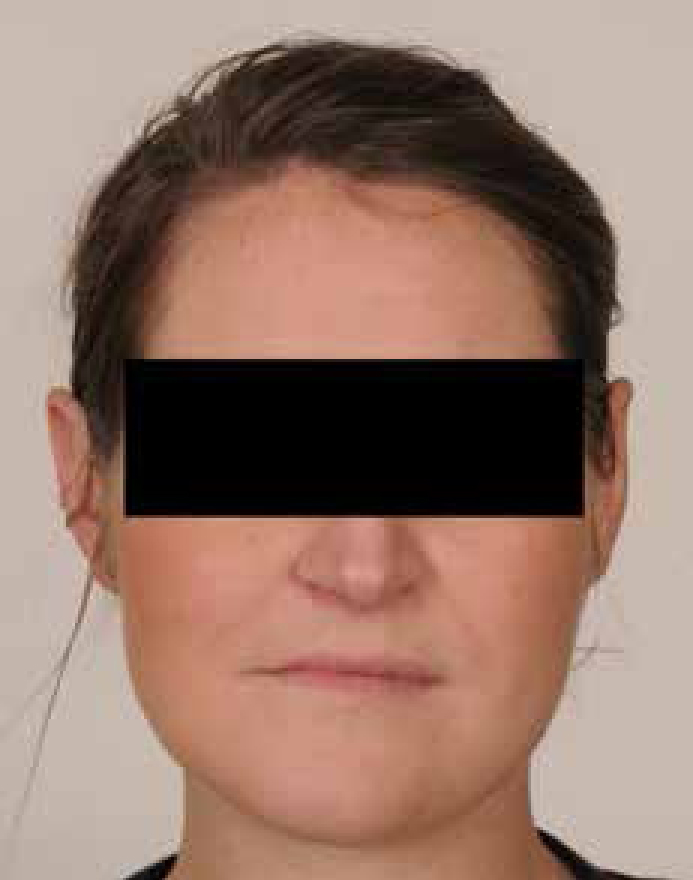
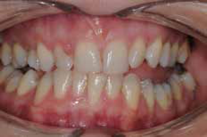
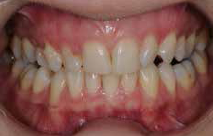
Clinical examination revealed enlargement of the left mandibular condyle in both lateral and posterior dimensions, and the left gonial notch more inferiorly placed when compared to the right. An element of bowing of the lower border of the mandible was noted, with deviation of her chin point to the right (Figure 1). Dental examination revealed an inter-incisal opening of 34 mm, and an obvious left-sided open bite on a Class III skeletal base (Figure 2).
Figure 1. Extra-oral view showing the obvious facial asymmetry.Figure 2. Pre-operative intra-oral views demonstrating a unilateral left-sided open bite.
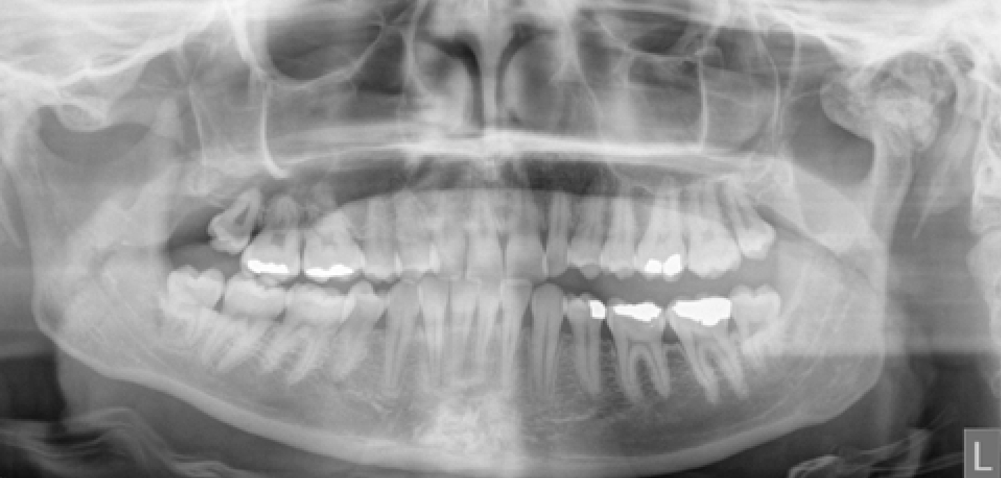
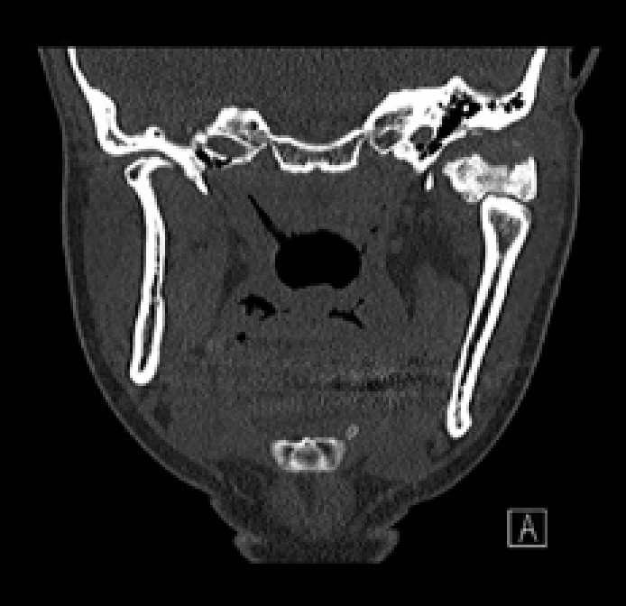
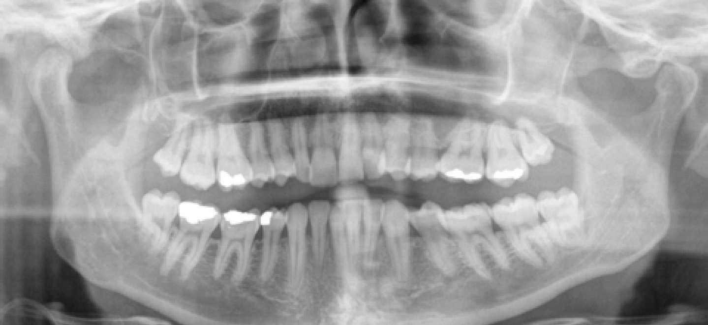
A panoramic radiograph was performed showing a bony outgrowth with heterogeneous radio-opacity extending medially from the left mandibular condyle (Figure 3). Subsequent CT demonstrated a bony mass present between the head of the left condyle and the glenoid fossa, continuous with the anterior aspect of the condylar head, with a maximum antero-posterior dimension of 4 cm (Figure 4). The lesion was largely smooth in outline apart from one anterior area consistent with a pseudoarthrosis. No union between the condyle and the glenoid fossa was observed, although the fossa was irregular in outline. The cortex and medulla of the lesion were continuous with the same structures of the mandibular condyle.
Figure 3. OPG demonstrating the bony outgrowth of heterogeneous radiographic density extending medially from the left mandibular condyle.Figure 4. Coronal CT demonstrating a bony mass present between the head of the left condyle and the glenoid fossa.
Excision of the lesion with condylar remodelling and disc repositioning was performed under general anaesthetic via an extended temporal/pre-auricular approach. Microscopically, sections showed areas of normal bony appearance surrounded by a cap of hyaline cartilage. The features described are those of an osteochondroma.
Discussion
Osteochondromas are cartilage-capped exophytic lesions that arise from the cortex of bone. They constitute between 10–15% of all bone tumours.4 They most commonly develop in the long bones, especially the distal metaphysis of the femur and proximal metaphysis of the tibia.
Osteochondromas rarely affect the craniofacial region. When they affect the facial skeleton the most common sites are the medial surface of the coronoid process of the mandible and the mandibular condyle.5,6
The majority of reported cases present in the fifth decade, predominantly in females, and involve the mandibular condyle.5 There are no large studies available for review with most of the literature consisting of single case reports.
Various aetiologies have been proposed for osteochondromas, including the persistence of areas of immature chondrocytes in the periosteum.6 These areas may be acted upon by mechanical stress, and lead to hyperplasia. This would be in line with the common occurrence of these lesions, usually located in areas of tendon insertions. There has also been a suggestion that remnants of Meckel's cartilage play a role in these lesions.7 Sarcomatous transformation in osteochondroma is reported to occur in 2% of lesions. To the best of our knowledge, no case has been reported in a craniofacial lesion.
Resection is often complex and requires a combination of approaches and methods. Such procedures are deemed curative, with only one recurrence of a condylar osteochondroma reported in the literature. This occurred one year after excision of a fragmented lesion.8
Whilst osteochondroma of the condyle is rare, it is a condition that could present to the general dental practitioner. Symptoms and signs that should alert the practitioner to the possible diagnosis are:
Malocclusion;
Lateral open bite on the affected side with contralateral cross-bite;
Osteochondromas are found at various sites around the condyle. CT and MRI scans are mandatory to establish accurate diagnosis and to plan the approach to treatment.12 Treatment must be undertaken at a specialist maxillofacial unit.
The majority of reported cases have been treated by a sub-condylar resection of the lesion with or without reconstruction.13 Post condylectomy, a loss of ramal height, can result in the formation of an open bite. Condylar reconstruction is often advocated to decrease postoperative occlusal difficulties.14 Reconstruction of the condyle can be accomplished by free autogenous bone graft, osteotomy, or a joint prosthesis. These procedures, however, can increase both the time and complexity of the operation and add to possible post-operative complications.
In this case, the mass was removed, the remaining head of condyle re-contoured and the disc replaced. This has the advantage of maximizing ramal height, and reducing the need for a more complex procedure. As this procedure has the theoretical disadvantage of increased recurrence, regular clinical and radiographical follow-up is necessary.
The post-operative course was generally uneventful. Review at one year confirmed good mandibular movement, with inter-incisal opening of 44 mm and normal facial nerve function. The patient was encouraged to continue to exercise the joint gently to prevent any fibrous scarring of the area. Her occlusion was much improved, with bilateral posterior molar contacts, and a residual minimal anterior open bite. Despite post-operative inter-maxillary elastic therapy, there remains a half unit mandibular centre line shift to the right (Figure 5). OPT revealed no signs of recurrence of the lesion, or change in size of the residual condylar head (Figure 6).
Figure 5. Post-operative intra-oral views with bilateral molar contacts at 3 months.Figure 6. Post-operative OPG at 3 months.
Mandibular and occlusal asymmetries can have many causes, and often present first to the general dental practitioner. In our opinion it was the urgent referral for appropriate investigation and treatment to a specialist unit that lead to an uncomplicated, positive outcome for our patient.