Erten H, Uctasli MB, Akarslan ZZ The assessment of unaided visual examination, intraoral camera and operating microscope for the detection of occlusal caries lesions. Oper Dent. 2005; 30:(2)190-194
Kidd EAMOxford: Oxford University Press; 1996
Sitbon Y, Attathom T, St-Georges AJ Minimal intervention dentistry II: Part 1. Contribution of the operating microscope to dentistry. Br Dent J. 2014; 216:(3)125-130
Clark DJ, Sheets CG, Paquette JM Definitive diagnosis of early enamel and dentin cracks based on microscopic evaluation. J Esthet Restor Dent. 2003; 15:(7)391-401
Perrin P, Jacky D, Hotz P The operating microscope in dental practice: minimally invasive restorations. Schweiz Monatsschr Zahnmed. 2002; 112:(7)722-732
Bonsor SJ Contemporary use of flowable resin composite materials. Dent Update. 2008; 35:(9)600-606
Jurkschat U Improvement of stomatological diagnosis by means of the II operating microscope. Stomatol DDR. 1979; 29:(1)15-18
Clark D The operating microscope and ultrasonics; a perfect marriage. Dent Today. 2004; 23:(6)74-81
Nase JB The clinical operating microscope advantage in fixed prosthodontics. Gen Dent. 2003; 51:(5)417-422
Friedman M, Mora AF, Schmidt R Microscope-assisted precision dentistry. Compend Contin Educ Dent. 1999; 20:(8)723-736
Carr GB, Murgel CA The use of the operating microscope in endodontics. Dent Clin North Am. 2010; 54:(2)191-214
Khayat BG The use of magnification in endodontic therapy: the operating microscope. Pract Periodontics Aesthet Dent. 1998; 10:(1)137-144
Saunders WP, Saunders EM Conventional endodontics and the operating microscope. Dent Clin North Am. 1997; 41:(3)415-428
Song M, Kim HC, Lee W, Kim E Analysis of the cause of failure in nonsurgical endodontic treatment by microscopic inspection during endodontic microsurgery. J Endod. 2011; 37:(11)1516-1519
Wu D, Shi W, Wu J The clinical treatment of complicated root canal therapy with the aid of a dental operating microscope. Int Dent J. 2011; 61:(5)261-266
Evans GE, Bishop K, Renton T Update of guidelines for surgical endodontics – the position after ten years. Br Dent J. 2012; 212:(10)497-498
Kersten DD, Mines P, Sweet M Use of the microscope in endodontics: results of a questionnaire. J Endod. 2008; 34:(7)804-807
Buhrley LJ, Barrows MJ, Begole EA, Wenckus CS Effect of magnification on locating the MB2 canal in maxillary molars. J Endod. 2002; 28:(4)324-327
De Carvalho MC, Zuolo ML Orifice locating with a microscope. J Endod. 2000; 26:(9)532-534
Karapinar-Kazandag M, Basrani BR, Friedman S The operating microscope enhances detection and negotiation of accessory mesial canals in mandibular molars. J Endod. 2010; 36:(8)1289-1294
Schwarze T, Baethge C, Stecher T, Geurtsen W Identification of second canals in the mesiobuccal root of maxillary first and second molars using magnifying loupes or an operating microscope. Aust Endod J. 2002; 28:(2)57-60
Stropko JJ Canal morphology of maxillary molars: clinical observations of canal configurations. J Endod. 1999; 25:(6)446-450
Gorduysus MO, Gorduysus M, Friedman S Operating microscope improves negotiation of second mesiobuccal canals in maxillary molars. J Endod. 2001; 27:(11)683-686
Biswas M, Mazumdar D, Neyogi A Non surgical perforation repair by mineral trioxide aggregate under dental operating microscope. J Conserv Dent. 2011; 14:(1)83-85
Kahler B Microsurgical endodontic retreatment of a maxillary molar with a separated file: a case report. Aust Dent J. 2011; 56:(1)76-81
Gencoglu N, Helvacioglu D Comparison of the different techniques to remove fractured endodontic instruments from root canal systems. Eur J Dent. 2009; 3:(2)90-95
Saunders WP, Saunders EM Coronal leakage as a cause of failure in root-canal therapy: a review. Endod Dent Traumatol. 1994; 10:(3)105-108
Saunders WP, Saunders EM Assessment of leakage in the restored pulp chamber of endodontically treated multirooted teeth. Int Endod J. 1990; 23:(1)28-33
Kim S Principles of endodontic microsurgery. Dent Clin North Am. 1997; 41:(3)481-497
Pecora G, Andreana S Use of dental operating microscope in endodontic surgery. Oral Surg Oral Med Oral Pathol. 1993; 75:(6)751-758
Tibbetts LS, Shanelec DA A review of the principles and practice of periodontal microsurgery. Tex Dent J. 2007; 124:(2)188-204
Setzer FC, Shah SB, Kohli MR Outcome of endodontic surgery: a meta-analysis of the literature – Part 1: Comparison of traditional root-end surgery and endodontic microsurgery. J Endod. 2010; 36:(11)1757-1765
Setzer FC, Kohli MR, Shah SB Outcome of endodontic surgery: a meta-analysis of the literature – Part 2: Comparison of endodontic microsurgical techniques with and without the use of higher magnification. J Endod. 2012; 38:(1)1-10
Shanelec DA Anterior esthetic implants; microsurgical placement in extraction sockets with immediate provisionals. CDA Journal. 2005; 33
Bowers DJ, Glickman GN, Solomon ES, He J Magnification's effect on endodontic fine motor skills. J Endod. 2010; 36:(7)1135-1138
The use of the operating microscope in general dental practice part 2: if you can see it, you can treat it! Stephen J Bonsor Dental Update 2024 42:1, 707-709.
Authors
Stephen JBonsor
BDS(Hons) MSc FHEA FDS RCPS(Glasg) FDFTEd FCGDent GDP
The Dental Practice, 21 Rubislaw Terrace, Aberdeen; Hon Senior Clinical Lecturer, Institute of Dentistry, University of Aberdeen; Online Tutor/Clinical Lecturer, University of Edinburgh, UK.
The use of magnification by dental clinicians when carrying out examinations and treatments is becoming more commonplace. The best instrument for this purpose is the operating microscope which has been shown to enhance quality, longevity and outcome of clinical work as well as facilitating better ergonomics for both the dentist and dental nurse. This paper, the second of two, explores the potential uses for the operating microscope in general dental as well as specialist practice (such as endodontics) and discusses how the interested clinician can use such equipment in a practical manner.
Clinical Relevance: The operating microscope enhances the dental surgeon's vision so improving treatment outcomes not only in specialist fields, such as endodontics, but also in many of the disciplines which general dental practice encompasses.
Article
This paper follows on from the first in this two part series which explained how the interested clinician may embark on his/her journey using an operating microscope in dental practice. The use of such an instrument has been shown to enhance quality, longevity and outcome of clinical work in many branches of surgery as well as dentistry. Both the dentist and dental nurse using a microscope have improved posture and work in a more ergonomic manner. At the chairside, close support (four-handed) dentistry is facilitated as the dental nurse is more involved in the procedure owing to improved vision and, as a result, enhanced job satisfaction and interest.
Dental operating microscopes have many different features which may be useful, depending on the scope of the clinician's practice. Photography and video are often used as an adjunct to the use of the microscope and this has many advantages. Clinical records are enhanced, with clinical images being available to monitor hard or soft tissue lesions or as medico-legal evidence. These images are also invaluable in patient education.
The use of microscopy demands that bespoke microsurgical instruments need to be used in conjunction with the microscope. Both this equipment and the microscope itself must be carefully cared for and maintained as the hardware is expensive.
This paper focuses on the specific uses of an operating microscope in dental practice and how it may be used to facilitate the work of the clinician with the aim of improving the quality of the treatment which the patient receives.
Examination and diagnosis
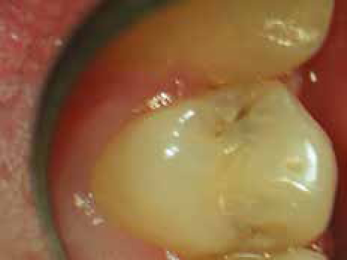
The operating microscope can greatly facilitate examination and diagnosis of the dental patient. This author has used an operating microscope to examine the oral cavity and its structures for over nine years. Pathologies such as dental caries1 (Figure 1), the presence of enamel infractions, and cracks in tooth structure and soft tissue lesions may all be more easily identified and examined by microscopy. That said, it is inadvisable only to use microscopy when doing an oral examination. This author prefers to examine all of the oral structures initially with the naked eye before proceeding to use the microscope. There are two reasons for this. First, this approach gives the dentist a ‘feel’ for the whole picture and therefore he/she is distracted by minutiae and secondly, suspicious areas or lesions may be noted mentally and then examined more thoroughly at a higher magnification. In practice, this has been well received by patients who are reassured that their mouth is being examined not once but twice during the examination appointment.
Figure 1. A small carious lesion in an upper premolar tooth which is more clearly seen at a higher magnification, in this case x8.
Examination and diagnosis of dental caries and cracked tooth tissue
Diagnosis of dental caries demands that the teeth are clean and dry to allow proper examination of their surfaces for lesions.2 The appearance of carious lesions changes when the teeth are dry so making them easier to visualize and examine. Small lesions may be treated conservatively (see later) and a better prognosis results with early intervention. Lesions noted without optical aids, which are suspected to be carious, may appear at higher magnification to be dark and shiny. In the absence of plaque, such lesions may be deemed to be inactive3 and should be documented in the clinical records. Such lesions should be kept under observation and reviewed at subsequent examinations. Without magnification the clinician may have been more inclined to intervene operatively.
For a dental examination, this author would recommend that the teeth are examined without microscopy in the first instance (as mentioned earlier) using a dental mirror, air from the 3 in 1 syringe and perhaps a dental probe (explorer) if necessary to remove debris only. This examination is then repeated under magnification using the air from the 3 in 1 syringe to dry the teeth being examined, as well as using it to retract the cheeks and lips to improve access and therefore vision. The operator works from quadrant to quadrant adjusting the position of the microscope as he/she proceeds so that each tooth is in sharp focus. Any suspicious carious lesions or possible defective margins of, or cracks and fractures in, dental restorations identified with the naked eye may then be further examined in more detail using magnification.
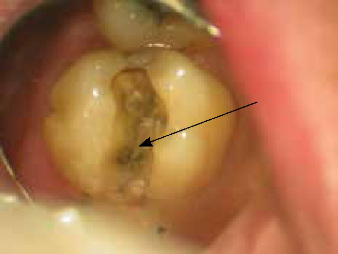
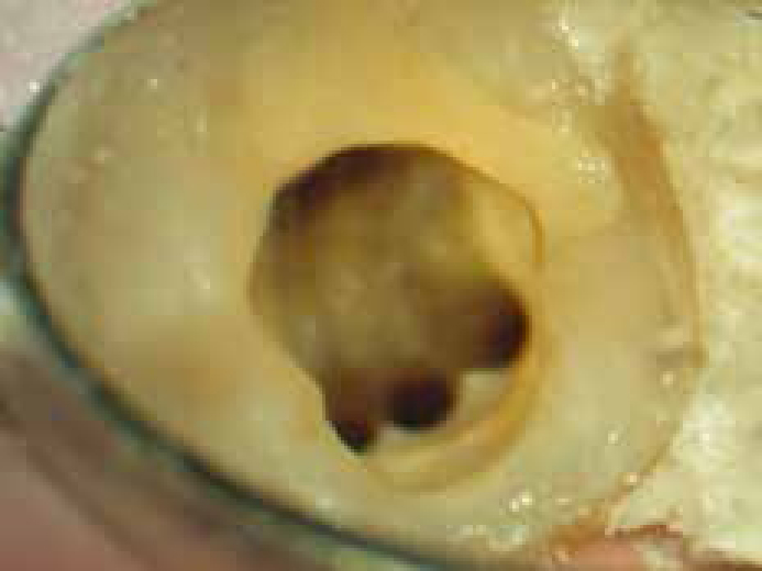
Microscopy has facilitated the identification and diagnosis of cracks in the dental hard tissues.4 This is best achieved by using a higher magnification such as a minimum of x13. Teeth may have fracture lines running at the base of the cavity floor (Figure 2) or under a cusp. Once a crack has been identified, a definitive diagnosis can be made and the treatment options may then be shared with the patient.4 Fractures are a leading cause of tooth loss today. The early diagnosis and treatment of cracked teeth and incomplete coronal fracture may prevent early loss of teeth. This treatment may involve the use of cuspal coverage using a bonding technique with either direct resin composite or dental amalgam or the provision of a cast restoration to brace and protect the remaining weakened tooth tissue from occlusal forces, so preventing crack propagation. The successful treatment of symptomatic incompletely fractured teeth is notoriously difficult and, with an uncertain outcome, as the aetiology of the condition is impossible to address, the aim of treatment is to mitigate the situation.
Figure 2. Fracture line extending from the distal marginal ridge on to the cavity floor (marked with an arrow).
Operative dentistry
Good vision is an advantage when administrating a local anaesthetic, a necessity as a precursor to many dental operative procedures. As the operator is more able to see the bevel on the needle and the mucosa, he/she may introduce it more precisely into the tissues, so reducing intra-injection discomfort. The advantages of this are obvious, particularly with nervous and trypanophobic (needle phobic) patients.
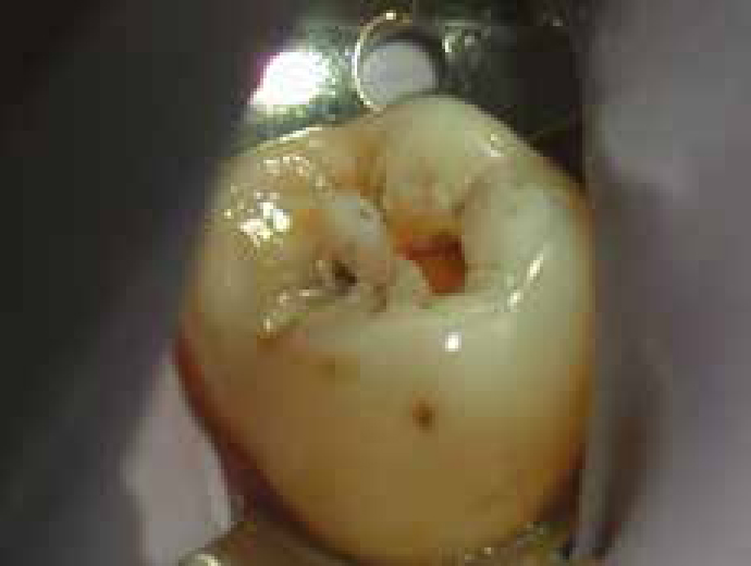
The operating microscope has revolutionized how conservative dentistry may be practised. The modern aim of conserving as much good tooth tissue as possible can only be done with the use of magnification.5 In recent years, there has been a move towards minimally invasive dentistry where a cavity may be prepared as conservatively as possible so preserving precious healthy tooth tissue (Figure 3). The cavity would then be restored with an appropriate dental restorative material such as a flowable resin composite.6 Microscopic techniques will facilitate this as both preparation and restoration are best done under magnification.3,7 The more tooth tissue that can be preserved the better, both in terms of operative dentistry and during endodontic preparation.8
Figure 3. The use of magnification facilitates the preparation of more conservative cavities such as this small lesion in a lower molar.
In fixed prosthodontics, magnification provides the opportunity of more precise tooth preparation for an indirect restoration.9 As the operator can see more clearly, better preparation geometry should result. This is especially so in areas which are more difficult to visualize clearly, such as approximal surfaces. The operating microscope can more easily provide an unimpeded view of these areas. Furthermore, the margins of indirect restorations may be placed more precisely in their desired position if the operator can see the area more clearly. This is best illustrated where a core material is present and the dentist wishes to site the crown preparation margin in its ideal position, usually apical to the margin of core material/tooth interface. At the fit appointment and subsequently at follow-up examinations, the margins of the cast may be more accurately assessed using microscopy. High magnification will also facilitate the removal of excess luting cement, resulting in better gingival health as no residual luting cement remains to provoke an inflammatory reaction in the gingival and periodontal tissues.
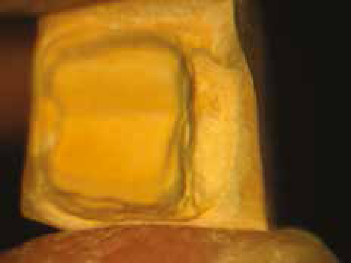
The microscope also has a role in the dental laboratory.10 The advantages of magnification may be extrapolated to the (more accurate) construction of dental appliances, especially in fixed prosthodontics. One example is the trimming of dies (Figure 4) which will be done with more precision under magnification, resulting in a better fitting indirect restoration.
Figure 4. The use of magnification facilitates the identification of the margins of the preparation on this die and allows more accurate trimming by the dental technician or clinician. Note that the occlusal surface of the preparation is slightly out of focus but the more apical crown margin is sharp. This demonstrates the importance of depth of field.
Conventional endodontics
Endodontists have embraced and benefited most from the increased magnification and illumination which an operating microscope provides.11,12,13,14,15 Most endodontists, or those dentists with a special interest in endodontics, use some method of magnification.16,17 In endodontics, the improved vision and illumination can facilitate:
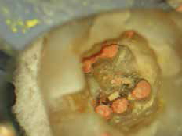
The identification and subsequent access of accessory anatomy18,19,20,21,22 (Figure 5);
The identification and removal of dystrophic calcification such as pulpal stones;
Improved quality of canal obturation and so ensuring the creation of an effective coronal seal (Figure 6);
The identification and subsequent repair of perforations and resorptive defects;24,25,26
The retrieval of separated endodontic instruments26,27 and fractured posts (especially non metallic posts which have been bonded in situ).
Figure 5. The use of the operating microscope facilities the identification of root canals such as a third canal in the mesial root of this lower left first mandibular molar.Figure 6. The same case as shown in Figure 5 after root canal obturation. The use of microscopy allows the endodontist to finish the obturation more precisely, so achieving a better coronal seal.
The examination of the tooth and the diagnosis of cracks have already been discussed and are significant in endodontics as these may be the cause of bacterial microleakage, leading to irreversible pulpitis, or may compromise the coronal seal, leading to failure of the endodontic treatment.28,29 Generally speaking, a higher magnification is used when performing endodontics than operative dentistry as the structures being managed are smaller.
Surgical endodontics and periodontics
The operating microscope is already used by many clinicians during periradicular30,31 and periodontal surgery.32 Setzer and co-workers have shown, in a meta-analysis of the literature, that outcomes of endodontic surgery are improved with the use of an operating microscope.33,34 The equipment allows microsurgical techniques to be employed. These techniques allow the soft tissues to be handled and replaced more accurately, which is associated with reduced surgical morbidity,16 as the tissues are more readily able to heal. Inaccurate apposition of the edges of the surgical flap, such as overlapping or invagination, will result in scarring. Increased magnification can at best eliminate and at worst reduce this as the surgeon may more accurately handle and control the final position of the tissues and suture the wound by passive primary closure. Consequently, as well as less scarring, less post-operative pain and more rapid healing will result. Microsurgical knots differ from conventional surgical knots as they have a ‘lumen’ which gives them the ability to compensate for any tightening of the tissues which is seen during initial healing due to oedema. Surgeons must learn these precision tailoring techniques at postgraduate courses and, once learned, it is advisable that they regularly practice knot tying to maintain their skills. This may be done by suturing a cut made in a piece of rubber dam.
The use of magnification allows a more conservative approach and the surgical site to be more easily controlled.30,31 This is particularly so in implant placement surgery as the fixture may be placed through a relatively small gingival incision, thereby minimizing tissue trauma and significantly reducing the post-operative pain. Healing and osseo-integration are both expedited and post-operative aesthetics improved.35 Other procedures associated with the placement of implants may also be facilitated with the use of an operating microscope, such as sinus lifts.
Minor oral surgery
It could be argued that the operating microscope has a more limited role in oral surgery. For example, exodontia would not be facilitated by the detail provided by a microscope. That said, this author has certainly used the operating microscope to examine sockets for the presence of retained roots or for the presence of oro-antral communications intra-operatively. The role of the operating microscope in periradicular and periodontal surgery has already been discussed and therefore the advantages of its use may be extrapolated to minor oral surgical procedures. Microscopy is already used for major oral and maxillofacial surgical procedures, such as the microsurgical connection of blood vessels and nerves in grafts after tumour resection.
Technique learning and development
When clinicians first begin to use an operating microscope they will find that the speed at which they work will be slower. There is a learning curve as they have to develop finer motor control and new ways of working.36 New techniques which are not possible without high magnification, such as microsurgical work and advanced endodontic techniques, will have to be learned and, to this end, attendance at postgraduate courses is to be highly recommended. However, with practice, most clinicians quickly become accustomed to working with the microscope.
The operating microscope is not useful in all branches of dentistry. Some subjects, such as orthodontics and removable prosthodontics, which are more concerned with ‘macroprocedures’, do not require such fine detail and so would not benefit from the use of a microscope.
Specialist society
In order for colleagues already using an operating microscope to interact professionally and to promote the advantages of its use, the European Society of Microscope Dentistry was founded in The Netherlands in 2007. This is an international association which encompasses all branches of microdentistry with its aims being:
To organize international microscope dentistry meetings on a regular basis in Europe;
To introduce the use of microscope magnification to all dental disciplines;
To provide initiating courses for those considering working with the microscope as well as courses for those wanting to enhance their skills in microscope dentistry;
To stimulate the implementation of the operating microscope in dental schools;
To encourage the development of techniques and equipment specifically for microscope dentistry;
To enhance open communication amongst microscope dentists.
The Society's website can be found at http://www.esmd.info
Conclusion
Once practitioners start to use magnification in their daily work, they find it impossible to return to operating without the help of optical aids. This is very much more the case with an operating microscope. The practice of clinical dentistry can be challenging but being able to see more clearly and being more comfortable at the chairside will make life much easier for the operator. Any dentist who has introduced an operating microscope into their clinical practice will surely testify that they are less fatigued at the end of the working day and the work they have produced is better. This has obvious benefits for the patient and increased job satisfaction for the clinician. Large teeth are easier to work on than small teeth and the use of the operating microscope will prove again and again that if you can see it, you can treat it!