Burke FJT Hemisection: A treatment option for the vertically split tooth. Dent Update. 1992; 19:8-12
Burke FJT, Crooks L Reconstruction of a hemisectioned tooth with an adhesive ceramic restoration using intraradicular retention. Dent Update. 1999; 26:448-452
Buhler H Survival rates of hemisection teeth: an attempt to compare them with survival rates of alloplastic implants. Int J Periodont Rest Dent. 1994; 4:(6)537-543
Stewart KL, Rudd KD, Kuebker WA Clinical Removable Partial Prosthodontics, 2nd edn. St Louis, Tokyo: Ishiyaku EuroAmerica Inc Publishers; 2003
Saad MN, Moreno J, Crawford C Hemisection as an alternative treatment for decayed multirooted terminal abutment: a case report. J Can Dent Assoc. 2009; 75:(5)387-390
Kryshtalskyj E Root amputation and hemisection. Indications, technique and restoration. J Can Dent Assoc. 1986; 52:(4)307-308
Green EN Hemisection and root amputation. J Am Dent Assoc. 1986; 112:511-518
Kost WJ, Stakiw JE Root amputation and hemisection. J Can Dent Assoc. 1991; 57:(1)42-45
Takei HH, Carranza FA The periodontal flap, 10th edn. In: Newman MG, Takei HH, Klokkvold PR, Carranza FA (eds). Missouri: Saunders; 2007
Newell DH The role of the prosthodontist in restoring root-resected molars: a study of 70 molar root resections. J Prosthet Dent. 1991; 65:(1)7-15
Prabhu NT, Munshi AK Hemisection of a permanent mandibular first molar: a treatment option for a vertically impacted second premolar. J Clin Pediatr Dent. 1996; 20:(3)233-235
The role of hemisection in the prosthetic management of a distal extension ridge – a case report AR Vivekananda Pai V Mohan Babu M Kundabala Dental Update 2024 41:6, 707-709.
Authors
AR VivekanandaPai
MDS
Professor and Head of the Department, Manipal College of Dental Sciences (Manipal University), Light House Hill Road, Mangalore – 575001, Karnataka, India
This case report illustrates the use of hemisection to minimize the distal extension span. The LR6 was the only molar next to a distal extension of the lower Kennedy's Class II ridge. Its unrestorable distal root was removed and its mesial portion was retained to serve as an effective antagonist and abutment tooth and lessen the extent of right distal extension.
Clinical Relevance: Regarding the prosthetic rehabilitation of distal extensions, hemisection can be advantageous and offered as an alternative to other treatment modalities.
Article
A distal extension removable prosthesis used for the rehabilitation of Kennedy's Class I and II ridges is associated with unfavourable leverage forces. The longer the edentulous span of distal extension, the greater will be force transmitted to the abutment teeth. Therefore, preservation of strategic terminal abutment teeth may be beneficial. In some situations, preserving a part of such a tooth may lessen the extent of distal extension span.
Hemisection is defined as ‘the division of a tooth in half and the removal of the unwanted, diseased portion, together with its root or roots’. It can be considered as a valid treatment since it follows the basic philosophy of conservative dentistry which is to aim to retain as much of the natural dentition as possible.1
Hemisection should be considered when the furcation of the molars is the result of either periodontal or non-periodontal problems like vertical root fracture. Further, it may be a valuable form of treatment when there is an extensive carious lesion extending subgingivally in one area of the root, making it impossible to place an adequate restoration in that area and making that root unrestorable.2
Studies regarding the success of hemisection have produced varying results. However, considering various criteria for failure, such as periodontal problems, endodontic complications, prosthetic problems, and caries, the average failure rate over a 7-year period was found to be 13.1%. Furthermore, the average failure rate was found to be 11%, when the number of cases in each study was taken into account.3 Despite these figures, the comparative lack of robust evidence means that hemisection should always be considered a ‘back to the wall’ treatment modality which may provide success in favourable conditions.
The removable prosthesis used for the rehabilitation of Kennedy's Class I and Class II ridges, which have bilateral and unilateral distal extensions, respectively, with limited treatment options, may be subjected to greater stresses as it is supported by soft tissue or a combination of tooth and soft tissue. Also, the distal extension denture base of the prosthesis can act as a lever arm and, therefore, the longer the distal extension span, the longer will be the denture base and the greater may be the force transmitted to the underlying ridge and abutment tooth. Hence, an increase in the extent of distal extension span should preferably be avoided by retaining as many teeth as possible. Moreover, from the point of view of efficient mastication, tooth retention rather than extraction and replacement is desirable. However, in certain situations, the retention of the whole tooth may not be possible. In certain circumstances, preserving a portion of the tooth, particularly of a molar, may be beneficial. For this purpose, given proper case selection and treatment planning, hemisection can be a viable option.4,5
The literature on the sectioning of a mandibular molar and the removal of its distal root is limited as this root is more often retained than the mesial root for anatomical reasons.2 Hemisection requires a multidisciplinary approach towards treatment, encompassing the realms of endodontics, periodontics and prosthodontics.6
This case report is about the hemisection of a distal root to meet the prosthetic treatment needs of the patient.
Case report
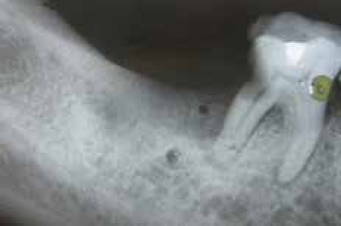
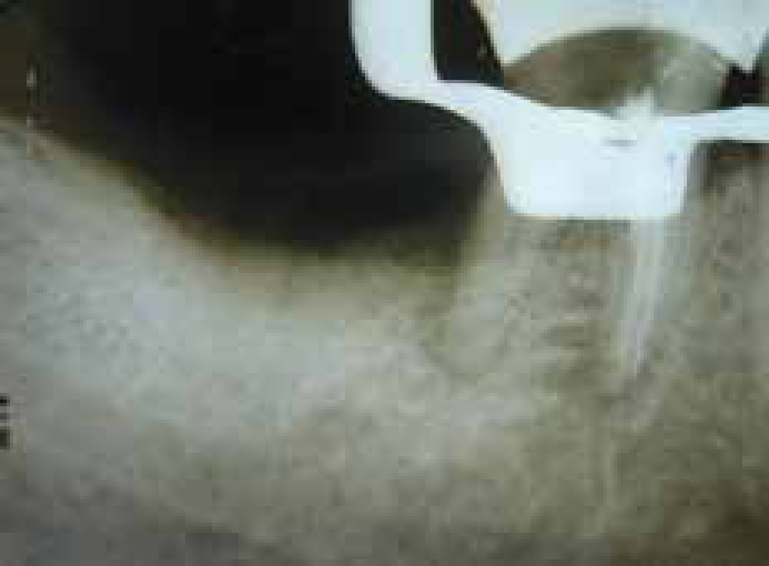
The patient, a 54-year-old woman, required the prosthetic rehabilitation of her upper and lower Kennedy's Class II, modification 1 type ridges (with unilateral distal extensions). It was planned to deliver a transitional, removable prosthesis to delay a tooth-tissue supported type of prosthesis for financial reasons. The patient was referred with severe pain in LR6 for three days. Clinical and periapical radiographic examinations of LR6 revealed an occlusal amalgam restoration with deep, subgingival distal proximal caries and 5–6 mm deep pocket on its disto-buccal aspect (Figure 1). As the tooth gave an exaggerated and prolonged response to the thermal and electric pulp tests and showed tenderness to percussion, along with apical widening of periodontal ligament space, a diagnosis of irreversible pulpitis with apical periodontitis was made and root canal treatment was suggested. Following treatment initiation and deep caries excavation, LR6 was deemed unrestorable at its distal aspect. However, its extraction was considered. LR6 was the only effective antagonistic tooth to UR5 and both were terminal abutments in their quadrants. Extraction of LR6 would have led to the loss of an antagonistic tooth and an increase in the extent of lower right distal extension. Therefore, it was decided to hemisect LR6 and retain its mesial half. Prior to hemisection, root canal therapy was performed in LR6 (Figure 2) and access opening was sealed using glass ionomer cement (GC Gold Label 2, GC Corporation, Japan).
Figure 1. Pre-operative radiograph of LR6.Figure 2. Radiograph following the obturation in LR6.
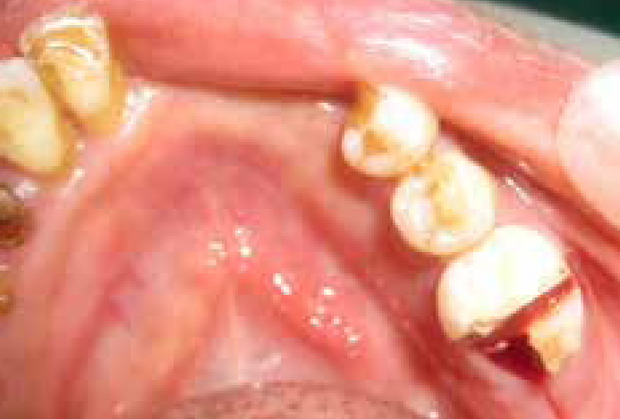
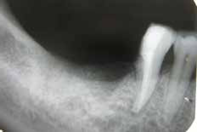
Hemisection of LR6 was performed under local anaesthesia, using an envelope flap design by giving only horizontal intrasulcular incision without any vertical component. The sectioning of the tooth was performed using a long diamond bur in a withdrawing motion, from the furcation to the crown, first mid-buccally, then mid-lingually (Figure 3). After confirming the complete separation, the distal half of the tooth was extracted (Figure 4). Following radiographic confirmation (Figure 5), the furcation side of the retained mesial half was contoured.
Figure 3. Occlusal view following the sectioning of LR6.Figure 4. Distal view following hemisection and extraction of distal root of LR6.Figure 5. Radiograph following the hemisection of LR6.
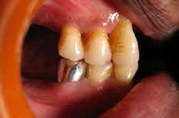
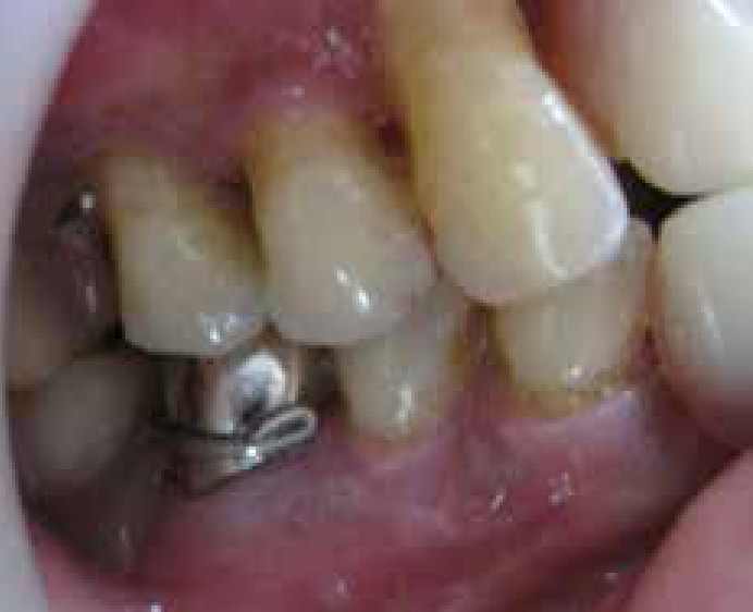
Following clinical and radiographic healing, the retained mesial half was restored with an all-metal crown to function like a premolar (Figure 6) and serve as an antagonistic and abutment tooth to engage the clasp extension of the tissue borne, acrylic-based transitional, removable prosthesis passively. At one year review, the prosthesis is well fitting and stable and the hemisected tooth is found to be asymptomatic and functional without any significant radiographic changes (Figure 7).
Figure 6. Hemisected LR6 with a metal crown functioning as an additional antagonistic premolar.Figure 7. One year follow-up view of hemisected LR6.
Discussion
Hemisection of LR6, in this patient, was beneficial as the extraction of LR6 could have led to the loss of a natural antagonistic tooth and an increase in the extent of the lower right distal extension, leading to reduced masticatory efficiency and the generation of greater stresses by the prosthesis, respectively. Furthermore, hemisection was suitable in this patient as only the distal portion of the LR6 was unrestorable and the mesial portion was intact.
Endodontic therapy was carried out prior to hemisection.7 The access opening was sealed with GIC, as materials such as amalgam are shown to affect the healing if lodged in the socket during hemisection.8 Since the periodontal defect was restricted to the disto-buccal portion of the root to be hemisected and flap displacement was not anticipated, an envelope flap design without vertical incisions was used. This flap ensures better adaptation and faster recovery with less post-operative complication.9 Tooth recontouring was required to ensure a smooth hemisected surface and eliminate residual furcal lips, which may go undetected and lead to periodontal problems.8,10
Although post placement is not routinely suggested in the retained root owing to certain inherent risks involved, a full crown is the preferred restoration as it prevents the fracture of the retained portion.7 The cuspal inclines of the crown should be minimized to control excessive lateral forces that could result in periodontal damage and occlusion should be carefully checked to balance the occlusal forces on the remaining root.5,6,10
For this patient, aesthetics was not of importance, so an all-metal crown was fabricated as a post endodontic restoration. Also, the metal crown would provide a better surface to engage and withstand the frictional forces generated during functioning of the prosthesis.
Many causes, like faulty resections, root fractures or endodontic complications, have been attributed to the failure of a hemisected tooth. The use of such a tooth as an abutment for multi-unit prosthetic reconstruction is another.11 Although presently, in this patient, the hemisected and crowned LR6 is passively engaging a tissue-supported transitional removable prosthesis, it may still be utilized as an abutment tooth for any future requirement of a tooth-tissue supported type of distal extension prosthesis, provided that the prosthesis can be designed to minimize undue stresses.
Conclusion
The removable prosthesis used for the rehabilitation of Kennedy's Class I and Class II type ridges leads to greater stress generation as the span of their distal extension increases. This can be minimized by retaining the tooth lying adjacent to the distal extension. However, when retention of the tooth is desirable, especially a multi-rooted molar, preserving a portion of it would be beneficial. Therefore, from a prosthetic treatment point of view, given a proper case selection and treatment planning, hemisection can be useful for preserving a portion of a tooth, particularly one acting as a natural antagonistic tooth, and it can be offered to the patient as an alternative to other treatment modalities.