Watts A, Addy M Tooth discolouration and staining: a review of the literature. Br Dent J. 2001; 190:309-316
Sulieman M An overview of tooth discoloration: extrinsic, intrinsic and internalized stains. Dent Update. 2005; 32:463-471
Kumar A, Kumar V, Singh J, Hooda A, Dutta S Drug-induced discoloration of teeth: an updated review. Clin Pediatr (Phila). 2012; 51:181-185
Nordbö H, Eriksen HM, Rölla G, Attramadal A, Solheim H Iron staining of the acquired enamel pellicle after exposure to tannic acid or chlorhexidine: preliminary report. Scand J Dent Res. 1982; 90:117-123
Waerhaug M, Gjermo P, Rølla G, Johansen JR Comparison of the effect of chlorhexidine and CuSO4 on plaque formation and development of gingivitis. J Clin Periodontol. 1984; 11:176-180
Vogel RI Intrinsic and extrinsic discoloration of the dentition. J Oral Med. 1975; 30:99-104
West NX, Addy M, Newcombe R A randomised crossover trial to compare the potential of stannous fluoride and essential oil mouth rinses to induce tooth and tongue staining. Clin Oral Investig. 2012; 16:821-826
Addy M, Roerts WR Comparison of the bisguanide antiseptics alexidine and chlorhexidine. J Clin Periodontol. 1981; 8:220-230
Bagis B, Baltacioglu E, Özcan M, Ustaomer S Evaluation of chlorhexidine gluconate mouthrinse-induced staining using a digital colorimeter: an in vivo study. Quintessence Int. 2011; 42:213-223
Addy M, Mahdavi SA, Loyn T Dietary staining in vitro by mouth rinses as a comparative measure of antiseptic activity and predictor of staining in vivo. J Dent. 1995; 23:95-99
Claydon N, Hunter L, Moran J A 6-month home usage trail of 0.1% and 0.2% delmopinol mouthwashes. Effects on plaque, gingivitis, supragingival calculus and tooth staining. J Clin Periodontol. 1996; 23:220-228
Addy M, Moran JM The formation of stain on acrylic surfaces by the interaction of cationic antiseptic mouth rinses and tea. J Biomed Mater Res. 1984; 18:631-641
Carpenter GH, Pramanik R, Proctor GB An in vitro model of chlorhexidine-induced tooth staining. J Periodontal Res. 2005; 40:225-230
Zanatta FB, Antoniazzi RP, Rösing CK Staining and calculus formation after 0.12% chlorhexidine rinses in plaque-free and plaque covered surfaces: a randomized trial. J Appl Oral Sci. 2010; 18:515-521
Ayaslioglu E, Erkek E, Oba AA, Cebecioğlu E Doxycycline-induced staining of permanent adult dentition. Aust Dent J. 2005; 50:273-275
Garcia-López M, Martinez-Blanco M, Martinez-Mir I, Palop V Amoxycillin-clavulanic acid-related tooth discoloration in children. Pediatrics. 2001; 108
Matson KL, Miller SE Tooth discoloration after treatment with linezolid. Pharmacotherapy. 2003; 23:682-685
Kumaraguru T, Flanagam SE, Greeley SA Tooth discoloration in patients with neonatal diabetes after transfer on to glibenclamide. Diabetes Care. 2009; 32:1428-1430
Sánchez AR, Rogers RS, Sheridan PJ Tetracycline and other tetracycline-derivative staining of the teeth and oral cavity. Int J Dermatol. 2004; 43:709-715
Mello H The mechanism of tetracycline staining in primary and permanent teeth. J Dent Child. 1967; 34:478-487
Jordan RE, Boksman L Conservative vital bleaching treatment of discolored dentition. Comp Cont Educ Dent. 1984; 5:803-808
Volovitz B, Shkap R, Amir J, Calderon S, Varsano I, Nussinovitch M Absence of tooth staining with doxycycline treatment in young children. Clin Pediatr (Phila). 2007; 46:121-126
Alvarez JA, Rezende KM, Marocho SM, Alves FB, Celiberti P, Ciamponi AL Dental fluorosis: exposure, prevention and management. Med Oral Patol Oral Cir Bucal. 2009; 14:E103-E107
Den Besten PK Mechanism and timing of fluoride effects on developing enamel. J Public Health Dent. 1999; 59:247-251
Pendrys DG, Katz RV, Morse DE Risk factors for enamel fluorosis in a nonfluoridated population. Am J Epidemiol. 1996; 143:808-815
Levy SM An update on fluorides and fluorosis. J Can Dent Assoc. 2003; 69:286-291
Lumbiganon P, Pengsaa K, Sookpranee T Ciprofloxacin in neonates and its possible adverse effect on the teeth. Pediatr Infect Dis J. 1991; 10:619-620
Auluck A, Thomas MS, Kundabala M, Sumanth KN Chlorodontia: minocycline induced staining of permanent dentition – a case report. J Conserv Dent. 2007; 10:89-92
McKenna BE, Lamey PJ, Kennedy JG, Bateson J Minocycline-induced staining of the adult permanent dentition: a review of the literature and report of a case. Dent Update. 1999; 26:160-162
Krastl G, Allgayer N, Lenherr P, Filippi A, Taneja P, Weiger R Tooth discoloration induced by endodontic materials: a literature review. Dent Traumatol. 2012; 29:2-7 https://doi.org/10.1111/j.1600-9657.2012.01141.x
Kim ST, Abbott P The effects of Ledermix paste as an intracanal medicament on the discolouration of teeth. Aust Endod J. 2000; 26:86-87
Kim JH, Kim Y, Shin SJ, Park JW, Jung IY Tooth discoloration of immature permanent incisor associated with triple antibiotic therapy: a case report. J Endod. 2010; 36:1086-1091
Tay FR, Mazzoni A, Pashley DH, Day TE, Ngoh EC, Breschi L Potential iatrogenic tetracycline staining of endodontically treated teeth via NaOCl/MTAD irrigation: a preliminary report. J Endod. 2006; 32:354-358
Krishnamurthy S, Sudhakaran S Evaluation and prevention of the precipitate formed on interaction between sodium hypochlorite and chlorhexidine. J Endod. 2010; 36:1154-1157
Hopcraft MS, Tan C Xerostomia: an update for clinicians. Aust Dent J. 2010; 55:238-244
Hellwig E, Lussi A Oral hygiene products and acidic medicines. Monogr Oral Sci. 2006; 20:112-118
Amaechi BT, Higham SM Dental erosion: possible approaches to prevention and control. J Dent. 2005; 33:243-252
Tooth discoloration is a common problem for which patients seek dental care. Various medications can directly or indirectly result in tooth discoloration. As clinicians, it is our responsibility to know these therapeutic drugs which can cause tooth discoloration and educate our fellow colleagues to take necessary precautions when prescribing these medications. Therefore, the objective of this paper is to give an overview of the various medications that can be linked to tooth discoloration and to suggest the precautionary measures that can be taken to avoid or minimize it.
Clinical Relevance: Dental discoloration potential of medications always needs to be considered before prescribing them.
Article
Discoloration of the tooth is a common problem for which patients seek dental care. Alteration in the tooth colour can not only cause aesthetic displeasure in individuals, but can also be traumatizing psychologically. Tooth discoloration can be either extrinsic (where chromogens are present on the surface of the tooth) or intrinsic (where chromogens are present within the bulk of the tooth structure) or even internalized (where external stains are taken up into the tooth surface through enamel defects and porous surface of exposed dentine).1,2,3,4 Among the several causes for tooth discoloration, a variety of medications used systemically or locally have been implicated in tooth dyschromia (Table 1).5 In the aesthetically conscious world of today, tooth discoloration that is iatrogenic can be a liability. The purpose of this article is to throw some light on the drugs that can either directly or indirectly result in tooth discoloration and the precautions that need to be taken when prescribing or using these medications.
Medications that reduce the salivary flow and its buffering capacity
Oral medications with sweetening agents
Medication-related dental erosion
Medications with low pH
Drugs that increase susceptibility to GORD
Medication-related primary tooth discoloration
Certain medications used locally or systemically are directly associated with extrinsic or intrinsic tooth discoloration. The prechromogens or the chromogens from these medications are either deposited on the surface or within the tooth structure to result in extrinsic and intrinsic discoloration, respectively.
Extrinsic discoloration
The medications that can stain the outer surface of the tooth, after it erupts into the oral cavity, can be of metallic or non-metallic origin.3 These superficial stains can usually be removed by oral prophylaxis.
Metallic stains
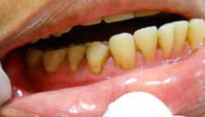
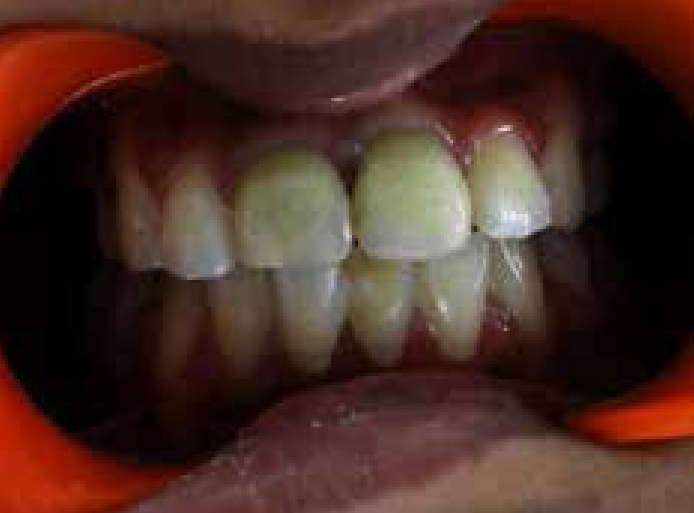
Oral solutions or mouthwashes containing metal salts: Medicaments containing metallic compounds have been associated with extrinsic staining of the teeth. For instance, dark brown to black discoloration can be seen in individuals consuming iron supplements (Figure 1).6 The use of mouthrinses containing copper salts can cause green staining on the teeth.7 Potassium permanganate mouthwash, which is used in patients with oral candidiasis and stannous fluoride mouthwash, which is used as a desensitizing agent, can cause violet-black and brown stains, respectively.8,9 The mechanism of staining of these metallic compounds is said to be because of the interaction of the metals with dental plaque. The sulphide salts of these metals were thought to cause the discoloration, but the exact mechanism of this chemical process is still unclear.3
Figure 1. Dark brown extrinsic staining due to iron containing medicament.
Non-metallic stains
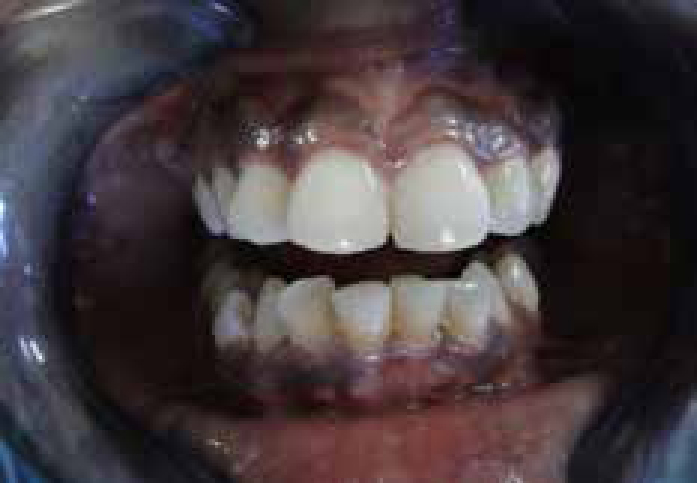
Mouthrinses: Extrinsic staining of the teeth can be a side-effect of certain antiseptic mouthwashes.10 The use of chlorhexidine, which is a cationic bisbiguanide, is shown to produce brownish discoloration in some individuals (Figure 2).11,12 Extended use of cationic quaternary ammonium compounds, like cetylpyridinium chloride,13 and other mouthwashes, like those based on phenolic essential oil7 and delmopinol hydrochloride,14 have also been linked with superficial tooth discoloration.
Figure 2. Brownish discoloration seen at the cervical area of lower incisors due to the extended use of chlorhexidine mouthwash.
The mechanism by which cationic mouthwashes produce extrinsic tooth staining is believed to be due to the precipitation of anionic dietary chromogens onto the adsorbed cations.15 Hence, it is advisable to ask patients on chlorhexidine mouthwash to reduce the consumption of beverages containing polyphenols (eg tea, coffee or wine) and rinse the mouth immediately after its consumption in order to decrease the occurrence of staining.16 Zanatta et al17 demonstrated that chlorhexidine staining was present significantly more in plaque-covered teeth than plaque-free teeth. Therefore, professional cleaning of the teeth to remove the plaque before prescribing chlorhexidine mouthwash can reduce the incidence of tooth staining
Systemic medications: Extrinsic tooth discoloration has also been noticed after the use of systemic antimicrobial agents like minocycline and doxycycline.18 They are thought to bind to the glycoprotein of the dental pellicle, especially in patients with poor oral hygiene. This is then said to undergo oxidation to produce discoloration when exposed to sunlight or bacteria. The above mentioned drugs have also been associated with intrinsic discoloration, the details of which will be discussed later in this paper. Systemic antimicrobial agents, like amoxicillin-clavulanic acid19 and linezolid,20 have also been linked with extrinsic discoloration. These drugs are said to cause pseudo-discoloration, more of which will be mentioned later. The use of glibenclamide to treat patients with permanent neonatal diabetes has also been shown to cause tooth discoloration.21 The cause of staining is said to be because of the precipitation of ingested chromogen onto the dental surface.
Intrinsic discoloration
Various medications have been implicated in intrinsic tooth discoloration, some of which may cause discoloration during odontogenesis (pre-eruptively) and others after odontogenesis (post-eruptively). Medications that cause discoloration during odontogenesis do it by changing the quality or quantity of enamel or dentine, or by incorporating the discolouring agent into the hard tissues. Medications that cause post-eruptive discoloration do it by incorporating the chromogens into the dental tissues, either through the tooth surface or pulp chamber.
Pre-eruptive discoloration
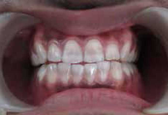
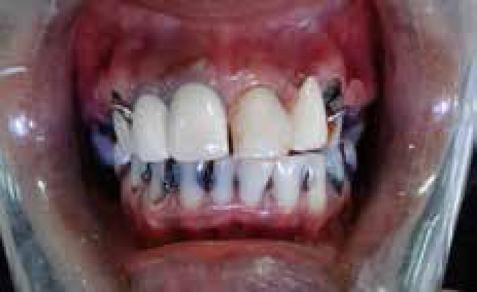
Tetracycline and its derivatives: These are broad spectrum antibiotics that are known to cause intrinsic tooth discoloration when prescribed during the formation of teeth (Figure 3).22 These antimicrobial agents are said to chelate with the calcium ions and thus form stable complexes in the hydroxyapatite crystals of enamel and dentine.23 The critical time during which tetracyclines can be incorporated into the deciduous anterior teeth is from 4 months in utero to 5 months post-partum and for the permanent dentition it is from 4 months post-partum to approximately 7 years of age. Therefore, tetracycline and its derivatives should be avoided in expectant and lactating mothers as well as children less than 8 years of age, unless absolutely necessary.3 Jordan and Boksman24 have classified tetracycline discoloration, based on the extent and degree of discoloration as:
First degree (light yellow, grey or brown, confined to incisal three-quarters with no banding);
Second degree (darker and more uniform discoloration without banding); and
Third degree (very dark blue or grey with banding).
Figure 3. Greyish discoloration caused due to pre-eruptive administration of tetracycline.
The severity and type of tooth discoloration depends on the type of tetracycline, its dosage, duration and period of exposure.4 The incidence of tetracycline-induced discoloration is shown to be high when administration of the drug is more than 3 g or when the duration of treatment exceeds 10 days.10
The type of discoloration is dependent on the type of tetracycline used. For instance, chlortetracycline produces slate grey discoloration, whereas tetracycline and oxytetracycline produce yellowish discoloration.1,3 A dental side-effect with the use of doxycycline has been shown to be less owing to its lower binding affinity for calcium.25 Minocycline, a semi-synthetic tetracycline (2nd generation tetracycline), unlike other tetracycline derivatives, has been shown to cause generalized greenish-blue discoloration post-eruption.26 The possible mechanism of this phenomenon will be discussed later. Tigecycline, a glycylcycline tetracycline derivative (3rd generation tetracycline), is also said to cause yellowish to brownish discoloration if administered during tooth development.27 Exposure to sunlight in tetracycline-affected teeth can change the colour to brown due to the photo-oxidative process. The affected teeth are also shown to give off a bright yellow colour under ultra-violet light due to fluorescence.3
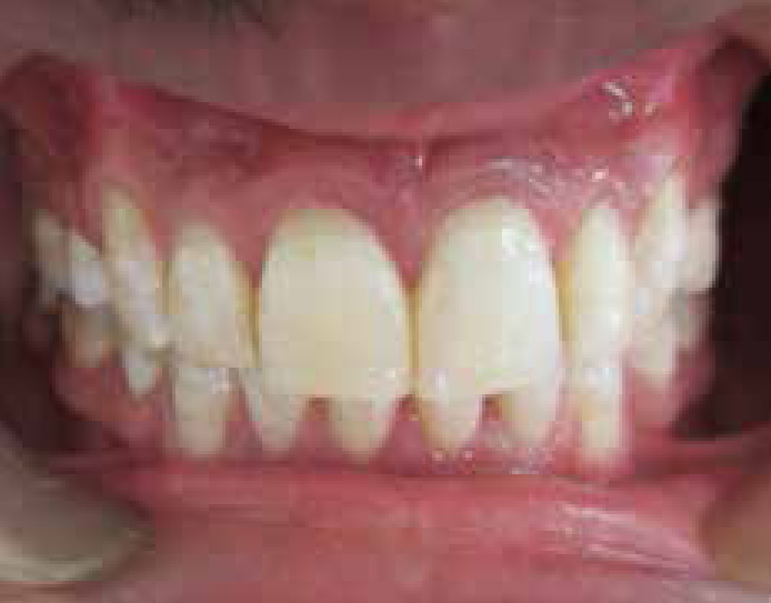
Fluoride supplements: Though fluorides have been shown to increase the resistance of the tooth to dental caries, its intake of more than the optimal level of 0.05–0.07 mg/kg body weight/day during the formative stage of the dental hard tissues can result in defective tooth mineralization, referred to as dental fluorosis (Figure 4).28 The enamel is frequently affected and the severity is dose dependent. In the mildest form it may appear as localized white flecks and in the severe form it may appear as diffuse opaque mottling with chalky white to dark brown colour and can even cause pitting of enamel.29 The dark discoloration is thought to be due to the internalization of extrinsic stains through the porous enamel.3 As the availability of fluorides can be from various sources, either natural (drinking water) or artificial sources (oral healthcare products, like fluoride mouthrinses, topical fluoride therapy, fluoridated toothpastes, fluoride tablets), it is the responsibility of the clinician to monitor the fluoride intake and judiciously use fluoride supplements as caries prevention means, especially in children during the critical period of teeth development (ie birth to 6 years of age).28,30,31
Figure 4. White opaque flecks seen in an individual from a non-endemic area that could be attributed to excessive fluoride from an artificial source.
Ciprofloxacin: Lumbiganon et al32 observed decalcification at the cervical part and greenish discoloration of the teeth which were resistant to mechanical removal in infants treated with 10 to 40 mg/kg/d ciprofloxacin for Klebsiella infection. Therefore, it was recommended by these clinicians to avoid the use of ciprofloxacin in newborn infants.
Post-eruptive discoloration
Medications that cause intrinsic discoloration in teeth erupted into the oral cavity usually enter the dental hard tissues through the pulp space and, in rare instances, through the intact enamel surface.
Minocycline: Minocycline is a semi-synthetic derivative of tetracycline that is commonly used in the treatment of acne vulgaris. Unlike other tetracyclines, minocycline is well absorbed from the gastrointestinal tract and its concentration in oral fluids is shown to be high.22 Extended use of more than 100 mg of this drug is shown to cause adult-onset greenish blue intrinsic discoloration of teeth in 3 to 6% of patients.10 This has been referred to as chlorodontia.33
Various theories have been suggested to explain the possible mechanism of minocycline introduced intrinsic staining which occurs post-eruptively in previously normal coloured fully mineralized adult teeth. One possible explanation for this is the ‘extrinsic theory’. According to this hypothesis, the oxidation of minocycline attached to the glycoprotein of the acquired pellicle results in the formation of insoluble black complexes. These pigments are then thought to be incorporated into the tooth structure by a demineralization-remineralization cycle, which is related to the high local levels of the drug.26,34 Another possible reason is the ‘intrinsic theory’, which suggests that minocycline gets deposited in collagen-rich tissues such as dentine during secondary or tertiary dentinogenesis. This is then slowly oxidized over time with the exposure to light.17,26 Yet another explanation for the mechanism of minocycline discoloration is by the ‘iron theory’, which assumes that hemosiderin, a product of iron metabolism, chelates with minocycline to form an insoluble complex resulting in discoloration.5,22
Intracanal medicaments: Various agents used to disinfect the root canal system have been linked with intrinsic discoloration. This is attributed by the internalization of stains through the dentinal tubules. Phenolic, iodoform and antibiotic-based medicaments that are sealed within the pulp space have been associated with tooth discoloration.35,36 Of these, it is the Ledermix paste (which contains triamcinolone acetonide and demethylchlortetracycline) and triple antibiotic paste (which contains ciprofloxacin, metronidazole and minocycline) that commonly cause tooth discoloration. The tetracycline derivatives present within the abovementioned medicaments are said to bind with the calcium of dentine-forming insoluble complexes, resulting in discoloration (Figure 5).37,38 Hence, alternative medicaments should be used, especially in teeth with greater aesthetic requirement.35 If these medicaments are to be used at all, they should be confined to the root canal apical to the gingival margin.36
Figure 5. Greenish discoloration of maxillary central incisors as a result of the placement of intra-canal medicament containing minocycline.
A few of the irrigants used during endodontic therapy have also been shown to cause discoloration, especially when used in combination with other irrigants. For example, when sodium hypochlorite (NaOCl) is combined with chlorhexidine, formation of a dark brown precipitate which is adherent to the walls of the pulp space is noticed.36 Even when BioPure MTAD (Dentsply, Tulsa, OK, USA)(mixture of a tetracycline, an acid and a detergent) was used as a final rinse after the use of NaOCl, formation of yellow precipitate along the root canal walls was observed. Photo-oxidation of the precipitate resulted in a red-purple tetracycline degradation product.39 If the abovementioned combination of irrigants has to be used, an in-between flushing of each irrigant with absolute alcohol/saline/distilled water and drying the canal before the use of the next irrigant should be carried out to prevent the formation of precipitates.40
Medication-related secondary tooth discoloration
Alteration of the oral environment or tooth structure can increase the susceptibility of the tooth to get discoloured. The oral environment may be altered by therapeutic drugs that can cause hyposalivation and those that can change the oral microflora. The therapeutic drugs that affect the tooth structure can increase the tendency of extrinsic stains to get internalized.
Alteration of the oral environment
Medication-related hyposalivation
As saliva helps to clear food particles and dental biofilm from the tooth surface, its reduction can result in the accumulation of debris, resulting in extrinsic staining. Medications that can result in the reduction of the salivary output (eg anticholinergics, antidepressants, anticonvulsants, etc) can thus indirectly be linked to staining.2,10 The importance of oral hygiene needs to be stressed in these patients.
Medication-related change in the oral microflora
Various antimicrobial agents (eg amoxicillin-clavulanic acid, linezolid) are shown to alter the oral microbial flora. An overgrowth of chromogenic micro-organisms can result in extrinsic pseudo-discoloration.10 These type of stains can be removed with oral prophylaxis.
Alteration of tooth structure
Medication-related dental caries
Medications that reduce the salivary flow and its buffering capacity can increase the susceptibility of the teeth to dental caries. Various oral medications with sweetening agents can also contribute to dental caries.10 In the initial stage, caries may appear as a white spot lesion and in the later stage it can appear as brown to black in colour owing to the adsorption of chromogens (Figure 6).3 Use of sugar-free gums, oral lubricants or even frequent sipping of water will give symptomatic relief in patients with dry mouth associated with drugs.41 Sugar-containing liquid medications should be avoided whenever possible.
Figure 6. Rampant caries associated with medication-induced hyposalivation.
Medication-related dental erosion
Certain medications with low pH (some mouthwashes, chewable aspirin, chewing hydrochloric acid tablets, dry powder antiasthmatic inhalers, etc) can cause direct loss of tooth structure due to erosion.42 Some therapeutic drugs may cause increased susceptibility to gastro-oesophageal reflex disease (GORD) (eg antiasthmatics, theophylline, anticholinergics, etc), thus leading to dental erosion indirectly.10 The loss of enamel may lead to yellowing of the teeth due to the exposure of underlying dentine. The porous surface left behind can also predispose the deposition and internalization of extrinsic stains.4 Patients should be advised to wash their mouth immediately after using these medications with neutral pH mouthrinses (tap water or neutral sodium fluoride mouthrinses) or basic mouthrinses (liquid antacids, sodium bicarbonate in water) to reduce the incidence of dental erosion.43
Conclusion
It is the responsibility of the clinician to warn the patient or the parent (in the case of a child) about the potential of a medication to discolour the teeth. Whenever possible, a safer alternative should be prescribed to minimize the incidence of staining. In situations where there are no alternatives, patients should be advised about the precautionary measures to minimize the stains. Those patients with discoloured teeth should consult a dental or medical practitioner to identify the aetiology and accordingly to consider the need for oral prophylaxis, dental bleaching, a restorative procedure or a combination of all three treatments.